Author Archives: Ken Ekechukwu

Simultaneous stenting of ostial stenosis of the common iliac arteries. Ken U. Ekechukwu, MD, MPH, FACP.

Smoking, dyslipidemias (disorders of lipids such as hypercholesterolemia), hypertension, and diabetes, individually and severally, may narrow or occlude arteries in all parts of the body.

Amongst arteries that may suffer are the ones that carry blood from the abdominal aorta (the large artery that carries blood from the heart across the abdomen towards the lower limbs), through the pelvis (the waist area), into the lower limbs, called the iliac arteries. If the narrowing of the vessel lumen is significant or the occlusion total, symptoms like claudication (pain during ambulation), paraesthesia, numbness, and erectile dysfunction may develop and bring a patient to medical attention.

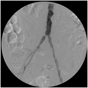
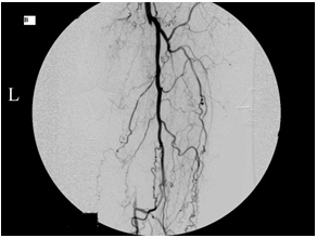
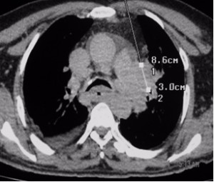
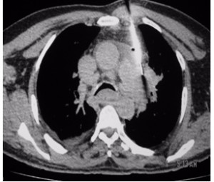
The images below illustrate such a problem. This patient in addition to having irregular chunks of calcium narrow his aorta has significant narrowing of the origins of his common iliac arteries (ostial stenosis), which along with the narrowings above and below them diminished the amount of blood reaching the patient’s feet from the aorta, causing symptoms.

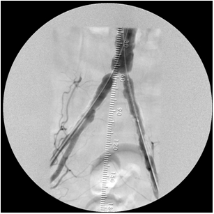
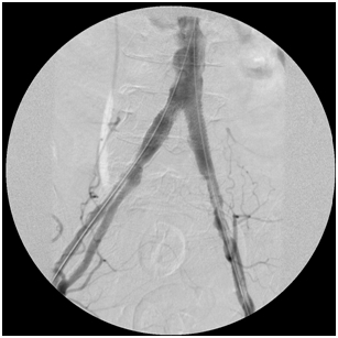
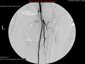
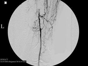
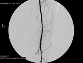
By simultaneously deploying a stent across each common iliac ostial narrowing, I corrected the problem, delivering more blood into the patient’s feet. The first image shows the narrowings before the stents were deployed; the second image displays the process of stent deployment; the third image was taken after I landed the stents.

Stent_deployment1 Stent_deployment2 Stent_deployment3


Treating arterial pseudoaneurysm by percutaneous injection of soluble thrombin. Ken U. Ekechukwu, MD, MPH, FACP.

An arterial pseudoaneurysm is a false sac that develops adjacent to an artery under its pulsatile influence, often after trauma to the artery. This includes arterial punctures. The wall of a pseudoaneurysm is not true (which is why it is called a false (or pseudo) aneurysm), being comprised of blood clot and compressed tissue, which explains why pseudoaneurysms may expand with time and cause symptoms.

When a pseudoaneurysm is diagnosed one of five treatment options are available: doing nothing, if the aneurysm is small; applying sustained pressure over its neck (that is the track connecting it to the native vessel) until it closes, also if it is a small pseudoaneurysm; repair it surgically; embolizing it with coils; injecting soluble thrombin into it under real-time ultrasound guidance, if it is reachable. More and more people now choose the last option to treat large symptomatic pseudoaneurysms that complicate arterial punctures because it is quick, simple to perform, and relatively safe. 

The images below show how I occluded a pseudoaneurysm of the right common femoral artery in a woman who developed one after cardiac catheterization.

The top 2 images show turbulent blood flow in the sac of the pseudoaneurysm, which in the second top image is shown to have a short narrow neck (the blue color below the turbulent sac) demonstrated to connect it to the right common femoral artery. The absence of turbulence in the 2 bottom row images means that the sac is thrombosed; the bright red color behind the sac in the lower second image is the common femoral artery, proving that it was not occluded during thrombin injection into the sac.

Pseudoaneurysm_pretx and posttx

Restoring blood flow across chronic total occlusion of the LSFA. Ken U. Ekechukwu, MD, MPH, FACP.

Fresh blood from the heart passes to the organs of the body through a large trunk of artery called the aorta that at about the level of the umbilicus divides into two equal arteries that cross the pelvis into the lower limbs. Each of the arteries in the lower limbs divides into two arteries in the upper thigh: one is the superficial femoral artery (SFA), which carries blood into the leg and foot past the knee, while the other, the deep femoral artery, supplies the thigh muscles.

If the superficial femoral artery is occluded or narrowed, cramps may develop in the leg muscles during walking or running. It usually begins mildly (although acute blockage will cause instant pain) as pain after walking a few blocks and worsens into pain after walking less than one block – if the stenosis or occlusion is not relieved. Much depends on the state of the arteries above and below the SFA.

This 75-year-old man’s primary care doctor referred him to my clinic because of left leg cramps after ambulating a few blocks and I found that his left superficial femoral artery was occluded (left column images). Using catheters, wires, balloons, and a covered stent, I restored blood flow through the blocked artery and his problem resolved (right column images).

Left_SFA_2 Left_SFA_6
Left_SFA_4 Left_SFA_5

Percutaneous biopsy of vertebral metastasis. Ken U. Ekechukwu, MD, MPH, FACP.

Sometimes it is necessary to biopsy a vertebra with metastatic disease for histopathology as in this woman in whom the T8 was the only evident site of destructive disease. Her primary site of cancer was unknown.

The approach to the percutaneous biopsy in this patient is not very different from the approach during vertebroplasty for thoracic vertebral compression fractures. Vertebroplasty is an effective method of relieving back pain in patients who have vertebral compression fractures.

 vertebral_metastatic_disease

Percutaneous biopsy of a lung (left hilar) mass. Ken U. Ekechukwu, MD, MPH, FACP.

Factors that determine whether pneumothorax will complicate percutaneous biopsy of a lung lesion are the size of the lesion, its depth in the lung, the size of the biopsy needle, and the number of needle passes during the biopsy. Fine needle aspiration (FNA) is safer than core biopsy because the needle size is smaller, but it is limited by the its inability to allow histopathologic tissue analysis and the need to have a cytopathologist present during the procedure to review specimens and declare their adequacy.

Lesions close to the hilum of the lung like in this case are best biopsied at bronchoscopy. In this case, however, the specimen obtained at bronchoscopy was inconclusive thus forcing a percutaneous biopsy that was only complicated by a small left pneumothorax. I observed the patient overnight in the hospital using periodic chest radiographs and no additional intervention was necessary.

Hilar masses_1 Hilar masses_2