History: An 87-year-old man was admitted to a hospital because of jaundice. He was found to have conjugated and unconjugated hyperbilirubinemia, the former more than the latter. His cross-sectional imaging revealed marked dilation of his extra-hepatic and intra-hepatic bile ducts that proved inaccessible at endoscopic retrograde cholangiopancreatography (ERCP). He was referred to interventional radiology for percutaneous biliary drainage, where the decision was made to percutaneously drain the biliary tree.
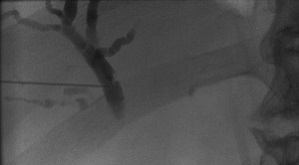
Intervention: Following adequate prophylactic antibiotic coverage against septicemia and under monitored-and-controlled anesthesia, a 22 gauge needle was blindly introduced into a branch of the right hepatic duct under fluoroscopy and confirmed by cholangiogram, Fig.1.
Fig. 1: Right hepatic cholangiogram through a 22 gauge needle introduced percutaneously from the right flank.

The needle was replaced with an AccuStick set over a guidewire through which a cholangiogram was repeated, revealing severe stricture of the distal right and left hepatic ducts as well as the common hepatic duct, Fig 2.
Fig. 2: Percutaneous cholangiogram through an AccuStick sheath in the proximal right hepatic duct.

Guide wires were advanced from accesses in the right flank and the epigastrium through the right and left hepatic ducts, respectively, past the common hepatic into the duodenum preparatory for inserting drainage catheters, Fig.3.
Fig. 3. Guide wires introduced through the hepatic ducts into the duodenum preparatory for catheter drainage.

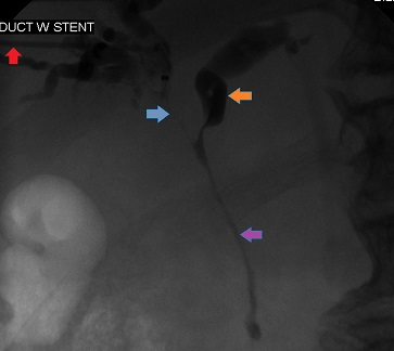
Balloons were passed over the guidewires to dilate the narrow tracts and an 8 French biliary drainage catheter was inserted into each tract. The cholangiograms through the catheters confirm optimal placement and marked decompression of the tracts, except the left hepatic duct, which still contained filling defects, Fig. 4. The patient did well after the procedure, but died before a histologic diagnosis was available. The final diagnosis was cholangiocarcinoma.
Fig.4. Post-procedure right and left cholangiograms through drainage catheters placed to decompress malignant biliary obstruction.


