Author Archives: Ken Ekechukwu

Juvenile Nasopharyngeal Angiofibroma Ken U. Ekechukwu, MD, MPH, FACP

History  
A 15-year-old boy saw his pediatrician for painless nontraumatic deformity of his left face and chronic nasal stuffiness. The pediatrician referred him to an ear, nose and throat (ENT) surgeon, who learnt from the boy’s mother that her son lost his ability to smell with his left nostril about 1 year before presenting and that he had had 2 episodes of epistaxis through the same nostril during that time. She had attributed his symptoms to common cold until his speech assumed a disturbing nasal quality and his left face swole 1 month before he presented. The surgeon noted a large mass in his left nasal fossa and biopsied it. Acting on the pathologic result of the biopsy specimen, he obtained maxillofacial cross-sectional imaging for the boy and referred him to an interventional radiology clinic for consultation.

Radiologic findings

ES2943_JNA_9 ES2943_JNA_1 ES2943_JNA_8
ES2943_JNA_5 ES2943_JNA_6 ES2943_JNA_7
ES2943_JNA_2 ES2943_JNA_3 ES2943_JNA_4
ES2943_JNA_11 ES2943_JNA_12 ES2943_JNA_10

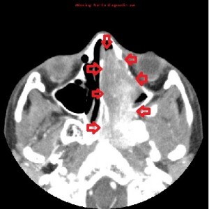
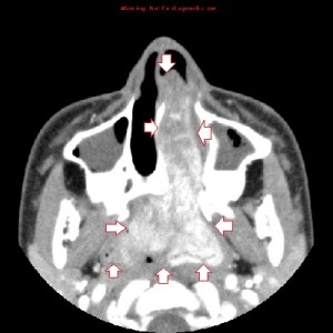
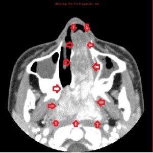
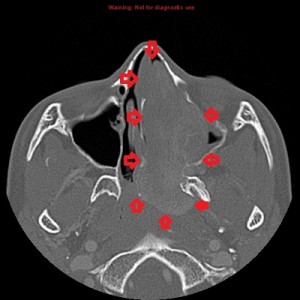
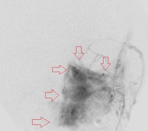
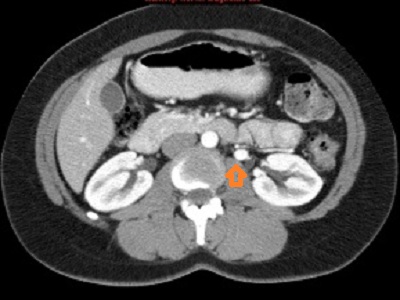
First panel, left to right: Axial contrast-enhanced maxillofacial CT scan in soft-tissue window from superior to inferior, showing a large, avidly enhancing mass in the left nasal fossa, extending posteriorly into the left nasopharnx and crossing into the right nasopharynx behind the nasal septum. Note opacification of the right and left maxillary sinuses, possibly from disturbed drainage.

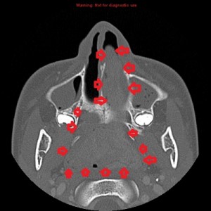
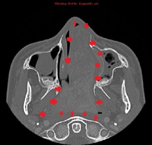
Second panel, left to right: The same set of images as in the first panel, but in bone window, showing bowing of the medial wall of the left maxillary sinus and opacified maxillary sinuses. There is no bone destruction or invasion.

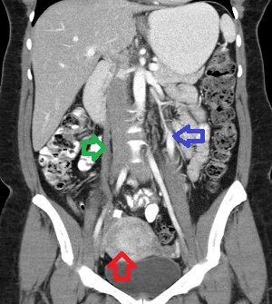
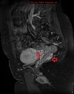
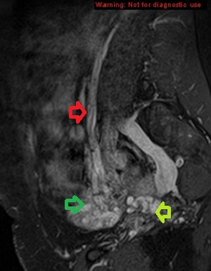
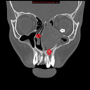
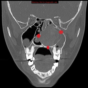
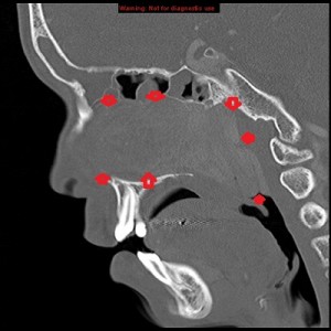
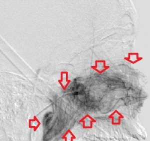
Third panel, left to right: The first 2 images are coronal sections of the face showing the mass-effect of the left nasal mass on other surrounding structure.The third image in this panel is a sagittal paramedian section showing obstruction of the nasopharynx by a soft-tissue mass.

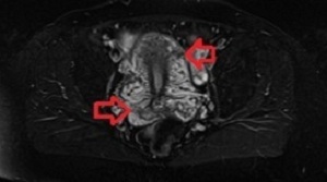
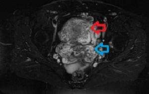
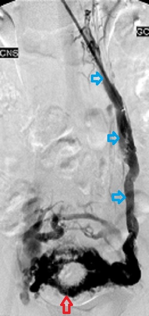
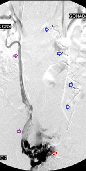
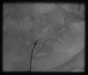
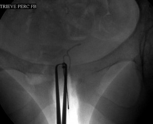
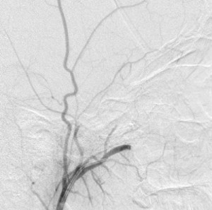
Fourth panel, left to right: Digital subtraction angiogram of the left internal maxillary artery before (1st two images, a frontal and a lateral view of the face) and after embolization (the last image, a lateral view). (For the 2nd and 3rd images, assume the patient’s face is turned to your right). Note the intense blushing of the tumor before embolization emphasizing its vascularity. Following embolization the mass is invisible.              

 Radiologic diagnosis: Juvenile Nasopharyngeal Angiofibroma.

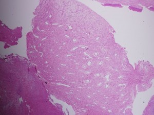
 Pathologic diagnosis: Juvenile Nasopharyngeal Angiofibroma.

OLYMPUS DIGITAL CAMERA OLYMPUS DIGITAL CAMERA OLYMPUS DIGITAL CAMERA

Discussion

Juvenile nasopharyngeal angiofibroma (JNA) is a benign locally aggressive hamartoma of the nasopharynx, first described in 1906 by Chaveau and constitutes only 0.05% of head and neck tumors. It occurs almost exclusively in the adolescent and young male population that its occurrence in a female should prompt chromosomal studies and measurement of serum 17-ketosteroids for sex determination. Its usual age range for occurrence is between 10 and 24 years, with a median age at diagnosis of 15 years.

JNA arises from the upper border of the sphenopalatine foramen and, as it grows, may extend into the infratemporal fossa, the sphenoid sinus, the maxillary antrum, the cavernous sinus, the middle cranial fossa , and, rarely, the anterior cranial fossa. Though its exact cause is unknown, the tumor is thought to arise from remnant  vascular plexus of the first branchial artery. Because it occurs at a time of facial bone development  50% of patients present with remodelled or damaged bones of the paranasal sinuses, more by expansion than by invasion. The most common bones affected are the maxilla, sphenoid, pterygoid plates, orbital apex and the skull base. In deed, the anterior bowing of the posterior wall of the maxilla at the level of the ptyregoid pillars, the Holman-Miller sign, gives the tumor its most consistent and reliable radiologic feature, although lymphoepithelioma and fibrous dysplasia are two rare nasopharyngeal tumors that have on occasion  caused anterior bowing of the posterior maxillary wall.

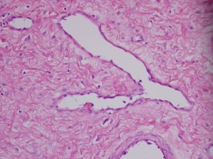
The tumor is composed of spindle-shaped  and stellate cells embedded in a collagen matrix and a complex of blood vessels that vary in size from capillaries to large venous channels and have no elastic lamina or elastic fibers; this latter characteristic explains its tendency to easy recurrent bleeding.  Thus,  severe recurrent epistaxis, occasionally life-threatening, occurs in 63% of patients, second only to unilateral nasal obstruction  (91%) as a presenting symptom. Other related symptoms of the disease are nasal discharge, pain, sinusitis, facial deformity, hearing impairment, otitis media, proptosis and diplopia, each reflecting JAN’s mass-effect on its neighbors. These symptoms generally present for 6 months to 1 year before the patient is diagnosed.

The diagnosis of JAN is straight forward: a young male with recurrent epistaxis and nasal obstruction found to have an obstructing nasal or nasopharyngeal mass in the absence of constitutional symptoms (fever, weight loss, lymphadenopathy (lypmhoepithelioma)). Standard imaging includes maxillofacial plain radiographs and contrast-enhanced computerized tomographic scan (CT scan) and magnetic resonance imaging (MRI).  The plain radiograph reveals a nasopharyngeal soft-tissue mass associated with anterior bowing of the posterior wall of the maxilla; CT  demonstrates the mass and any osseous involvement that may limit resection of the tumor and predict its recurrence; MRI’s superior soft-tissue imaging highlights the extent of the tumor and, with diffusion-weighted sequence helps distinguish JAN from a malignant nasopharyngeal tumor. Furthermore, MRI is useful for detecting tumor recurrence and should generally be obtained within 1 year after tumor resection for baseline information.

Severel treatment options for JNA have been described in the literature, some now obsolete. They include hormonal therapy (the tumor has receptors for many male hormones), radiation, embolization, chemotherapy, open surgical removal, and, in the last 2 decades, endoscopic resection of the mass. Hormonal therapy is largely abandoned for its ineffectiveness. Radiotherapy has been applied as definitive treatment for advanced JAN and has local control rates between 85% and 91%. However, it may be complicated by panhypopituitarism,  growth retardation, cataracts, radiation keratopathy, temporal lobe necrosis, and malignant transformation of the tumor, especially with high doses of radiation. It is perhaps excellent in treating primary or recurrent disease in critical areas, where surgical excision of the tumor may be hazardous. Open surgical extirpation was the only method of resecting the disease before the advent of modern novel techniques like endoscopic resection and radiofrequency coblation. There are many approaches to resecting the disease each dependent on the location and extent of the tumor. It is sometimes combined with endoscopic resection. Bland embolization of JNA is ususally offered as a presurgical intervention to limit hemorrhage during surgical removal. The usual arterial source to the tumor is the internal maxillary artery, but it may receive branches from the ophthalmic artery, ascending pharyngeal artery, and the facial artery. Identifying and embolizing these sources reduces the amount of intra-operative blood loss by 50% or more and creates a relatively bloodless operating field that improves tumor resection. When embolization is offered, surgical resection should be done in 2 to 5 days to avoid the development of collateral supply to the tumor. Endoscopic resection of the tumor enjoys certain advantages over traditional open surgery: comesis (no facial scars), shorter hospital stay that translates to lower cost; shorter recuperation; and better or equal recurrence rate. However, there are controversies over what stage of the disease is best treated by this technique. (I have eliminated the staging of JAN in this writing for want of space.)

Recurrence of JAN varies between 13% and 46% depending on the size of the tumor, its extension including osseous penetration, and the surgical approach used to remove it. Most recurrences occur within 12 months and are usually due to incomplete excision of the tumor.

This patient’s course

His laboratory investigation was unremarkable and the tumor was successfully resected endoscopically following presurgical embolization of his terminal left internal maxillary artery; the estimated blood loss was 350 mL. He did well following surgery but defaulted on followups. This month celebrates his first year of the resection of the tumor.

Acknowledgement    
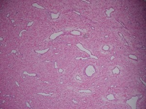
The pathology slides are by the kind auspices of Dr. Shanching Ying, Pathologist and Director of Microbiology Lab, Department of Pathology, Mount Sinai Medical Center, Chicago, Illinois.  


Gossypiboma Ken U. Ekechukwu, MD, MPH, FACP

History

A 50-year-old Mexican woman presented with abdominal pain to the emergency department of a hospital in the United States of America 12 months after a successful gastric surgery in her native land. She reported that 6 months after her surgery she developed epigastric discomfort and underwent treatment for what she was told was a suture granuloma. She did well for a short time but has had recurrent infection at the site marked by pain, a foul discharge from the site of surgery, subjective fever, nausea and vomiting – the same group of complaints that sent her to the emergency room this time. Except for hypothyroidism adequately treated with synthroid, past cesarean section and bilateral tubal ligation, her medical record was unremarkable. On examination, there was a dermal sinus in her epigastrium, bound by red inflamed tenderness and from which issued a foul discharge. She was afebrile and except for borderline leucocytosis, was fine. She had contrast-enhanced CT scan of the abdomen and latter an upper gastrointestinal series, the findings of which prompted a laparoscopic intervention. She did well afterward.

Radiologic findings

CT scan (top panel):
There is a round thick-walled mass ( arrowed red on all 3 CT images) anterior to the stomach, collapsing it. It has a spongiform matrix of bubbles of air mixed with unenhancing soft tissue and at its 8:00 position on the coronal view (the middle image), exhibits a radiodense non-metallic structure. Its 11:00 position on the axial view (the first image) is continuous with a midline thickening of the abdominal wall that proved to be a sinus at laparoscopy.

Upper gastrointestinal series (bottom panel):
There is a massive filling defect external to the stomach that markedly diminishes its capacity and corresponds in location to the mass seen on the CT scan. The red arrow on the first 2 images identifies radio-contrast in the collapsed stomach, the blue arrow identifies the extra-gastric mass, while the brown arrow identifies the radiopaque foreign body seen within the mass on the CT scan, which at surgery and pathology belonged to a surgical sponge. The 3rd image is a panoramic view of the upper midgut showing the mass, the stomach, and the proximal small bowel, demonstrating absence of gastric outlet obstruction. The topography of the gastric lumen is normal as shown on the double-contrast images (image not shown) and there is no communication between the mass and the stomach.

Surgico-Pathologic finding

The resected track of tissue between the mass and the skin at histology proved to be a fistula surrounded by “fibroadipose tissue and skeletal muscle with hemorrhage and focal chronic inflammation.” The mass was removed in fragments of friable foul tissue in the midst of which was a “blood-stained yellow gauze-like sponge measuring 16 cm x 10.9 cm x 3 cm.”

MC...4062 axial MC...4062 coronal MC...4062 sag

MC...4062UGI_1 MC...4062UGI_2 MC...4062UGI_3

Final diagnosis: Gossypiboma.

Discussion
 
Medical errors are common. Among them, though not as frequent as others but equally important, is the problem of unintentional retention of foreign objects in opened body cavities. Although our patient had her surgery outside USA, estimates suggest that every year in the USA  foreign objects are unintentionally left in 1500 patients and it is believed that these estimates may be low because such occurrences attract embarrassing press attention and punitive legal sanction that perpetrators are unwilling to report all. What is left in a patient varies: a surgical instrument  such as needle, staple, forceps, scissors, or a surgical sponge. The term gossypiboma describes a forgotten sponge and derives from Gossypium, Latin for cotton and boma, Swahili for a place of concealment. Textiloma is another term for lost surgical sponge since some surgical sponges are made from synthetic textile.
 
When a foreign object disappears in a patient there is a 50% chance that knowledge of the loss occurs within 3 weeks of surgery and 26% likelihood of such knowledge more than 2 months afterward. In all, 40% of gossypibomas are found within 1 year of their culprit surgery and 50% within 5 or more years. One of two things happens following unintentional retention of a foreign object in the body: an acute suppurative reaction may supervene as the body aggressively reacts to the foreigner or a more insidious accommodative granulomatous containment occurs as the foreign object is walled off into a calcified mass. The former is intense, with telltale features of acute infection – pain, fever, localized tenderness, diminished use of the affected area, etc – as the body attempts to destroy and expel the object. It may cause peritonitis, bowel perforation or obstruction, fistularization, septic shock syndrome and even death. The second form of reaction is relatively quiet and smoldering and may go undetected for many years. Walled-off foreign bodies have masqueraded as tumors in the body, being found serendipitously many years after they were forgotten. Our case exemplifies an intermediate type having smoldered for 12 months, its vigor blunted by intercurrent surgical interventions in Mexico and its vim diminished by the continuous emptying of its foul contents through an epigastric sinus.
 
There are other unsavory consequences of such error. In 69% of patients re-operation is inevitable. Legal adjudication is almost certain to incur a hefty penalty because negligence is obvious and its consequences on the patient self-evident. The press is gleeful in its exposition and decrying of wanton ineptitude and lack of care, attracting unwanted institutional public scrutiny and spot light with potential pocket book downsizing. The Centers for Medicare and Medicaid Services (CMS) currently does not pay for the treatment of errors caused by recent hospitalization, so the hospital will swallow the cost of clearing its mess – that is, if the patient returns to the original hospital. A visit from The Joint Commission on Accreditation of Health Care Organizations is every administrator’s nightmare. And, of course, there is the embarrassment to the surgeon responsible for the occurrence.
 
Foreign body retention can occur in any operation that involves opening of the body. Not surprisingly it does not occur with minimally invasive or laparoscopic surgeries. A majority occur with abdominal and gynecological surgeries with a few reported with cardiothoracic, neurosurgical, and other operations.
 
Research has shown that the following are risk factors or conditions that increase the likelihood of retaining a foreign body in a patient after surgery:
The urgency and nature of a procedure: Procedures performed emergently are 8.8 times more likely to be associated with retention of a foreign object in a patient than those done electively. Such procedures are often associated with speed and some measure of chaos that may lead to poor attention to instrument counting. In addition, procedures that are long, involve multiple operators or change of guards, or that are complicated have higher chance of being associated with foreign object retention. Under these circumstances, there is more blood loss and so more use of sponges and instruments with greater chance of losing some due to increase in number and reduced visibility from bleeding. Further, personnel may tire and be less attentive to detail and more likely to fail to fully communicate information to succeeding colleagues during shift changes. Thus, surgeons and nurses confronted with scenarios like these should be more vigilant to the possibility of unintentional retention of a foreign object and guard against it.

Unplanned change in a procedure: When there is an unscheduled change in the performance of a procedure, there is a 4.1 increased risk of leaving a foreign body behind in a patient. The reason is the confusion that such change may introduce to the procedure which makes foreign body retention more likely. In a sense it is an urgent or emergent surgery, not elective, and so prone to the vagaries of the former.

A patient’s body habitus: The size of a patient expressed as their body mass index has been shown to be associated with higher risk of postsurgical foreign body retention; the risk increases by 1.1 for each unit of increase in the patient’s body mass index. It is believed that a larger body mass provides more areas of concealment of surgical instruments and sponges and, to some degree, the tedium inherent in surgery on such patients may sap any remaining vigilance against losing foreign objects in them.

Poor counting discipline: Second only to not operating on a patient in the prevention of unintentional foreign body retention is the counting of surgical instruments and sponges. The importance of this exercise in reducing the risk of foreign body retention is underscored by the standards set for it by the Association of periOperative Room Nurses (AORN) – I refer the reader to the association’s web site, www.aorn.org, for details of its recommendations – and for this reason most preventive efforts aimed at unintentional foreign body retention have to do with instrument counting. Strict adherence to AORN’s standards significantly, but not completely, reduces the incidence of postoperative foreign body retention in patients. Disciplined accounting for every sponge or surgical instrument that makes a trip into a surgical field is the beginning of efforts to prevent leaving such in a patient.

Imaging problems: Most surgical instruments are radiopaque and so are easily identifiable when lost. Further, in USA since 1933, surgical sponges have been impregnated with barium in some form to enhance their visibility when lost. Thus, the principal mode of searching for a missing object in a patient in the operating room is radiography. But obtaining, interpreting, and transmitting the findings of a radiograph is not as seamless as it sounds. First, image acquisition is usually directed by the operating surgeon who may or may not be the final interpreter of the study. This can be problematic if the study fails to show the missing item and is interpreted by a radiologist unfamiliar with what the missing item looks like and who may be befuddled by the many surgical instruments that clutter the imaged surgical field. Clear instructions to the interpreting radiologist from the operating surgeon as to where and what to look for may help. Second, image acquisition may be suboptimal because it is portable and the operating field sterile and pregnant with many instruments; in addition the  patient may be obese, reducing x-ray penetration. Interpreting such poor-quality image either by the surgeon or a radiologist may be unrewarding. Attention to detail during image acquisition or the use of fluoroscopy under the direction of the surgeon or an invited radiologist may assist in this regard. It may be necessary to use CT scan ( I do not recommend other imaging modalities like sonography and MRI because they have less utility in this regard) to search for the missing object when the patient is out of the operating room. Third, the communication of the findings of a radiograph to the operating surgeon may be suboptimal especially if the study is interpreted after the patient is out of the operating room. Clear documentation of the findings of a study and the methods of communicating them to the requesting surgeon may help in this situation.
 
 
In summary, forgetting foreign objects in patients after surgery is a medical error with far-reaching consequences for the patient, health care providers, and health care institutions. Its avoidance requires combined individual vigilance and institutional supervision.

Pelvic congestion syndrome. U Ekechukwu, MD, MPH, FACP

History

 A 39-year-old parous woman presented to an emergency department with lower abdominal pain and CT scan of her abdomen was obtained as part of her investigation. Although the examination revealed no acute explanations for her symptom, it showed that her gonadal and pelvic veins were dilated, an observation present on a CT scan of her abdomen and pelvis obtained 3 years earlier. She was later referred to an interventional radiology clinic (IRC) for consultation for pelvic congestion syndrome and possible gonadal vein embolization.  Her medical history revealed a 6-year account of chronic abdominal pain, heavy menstrual periods, dysmenorrhrea and ultrasound-proven uterine fibroids. She had no gastrointestinal complaints except for diverticulosis without diverticulitis, nor did she have urological complaints or other gynecologic problems. There was no investigation of her psychological past nor were there any obvious manifeststions of its perturbation. She later had a pelvic MRI that did not confirm her past report of uterine fibroids but confirmed pelvic varices and bilateral gonadal vein dilation.

 She first underwent bilateral ovarian vein embolization with improvement in her symptoms that significantly improved after she underwent staged bilateral pelvic vein embolization several weeks later. She continues to do well and is still being followed at the IRC.

 Radiologic findings

First row, left to right: Axial (1st image) and coronal (2nd and 3rd images) contrast-enhanced MDCT scan of the abdomen and pelvis showing contrast opacification of the left ovarian vein in the arterial phase of the study (orange arrow on the 1st image and blue arrow on the second image) and unopacified but dilated right ovarian vein (green arrow on the 2nd image). There is beginning opacification of varicose uterine plexus arrowed red on the 3rd image.

Second row, left to right: Axial fat-suppressed T1WI of the pelvis of the same patient demonstrating dilated and tortuous uetrine and vaginal venous plexuses

Third row, left to right: Saggital fat-suppressed T1WI of the pelvis showing on the left image dilated left vaginal plexus and, on the right, dilated left ovarian vein (red arrow), dilated left ovarian plexus (blue arrow) and dilated pelvic veins (yellow).

Fourth row, left to right: First image, selective catheter left ovarian venogram showing dilated left ovarian vein (blue arrows), crossfilling of the contralateral pelvic veins through the uterine arcuate branches (red arrow). Second image, selective right ovarian venogram after embolization of the left ovarian vein confirming dilation of the vein (red arrows) and showing no opacification of the left pelvic veins across the uterine arcuates, proving successful embolization of the left ovarian vein (blue arrows). Third image, right and left ovarian veins after their embolization during right internal iliac venography.


Discussion

 Chronic pelvic pain in premenopausal women is common, being the reason for 10% of outpatient gynecologic visits. It is a symptom of several disorders that span multiple organ systems including gynecologic ( leiomyomas, endometriosis, adenomyosis, ovarian and tubal disorders, pelvic inflammatory disease), gastrointestinal (irritable bowel syndrome, inflammatory bowel disease) and urologic disorders. It is also a psychologic complaint in 40% to 60% of women with documented history of childhood sexual abuse and may be a somatization disorder. But when such pain occurs in a premenopausal woman, especially if multiparous, for 6 months or more in the presence of dilated ovarian or pelvic veins and has no explainable reason, it meets the definition of pelvic congestion syndrome (PCS). Thus, PCS is a disease of exclusion that requires multidisciplinary approach. The pain in pelvic congestion syndrome can be nonspecific, may occur with sexual intercourse, or PCS may manifest as persistent genital arousal or urinary urgency. The pain may be premenstrual, menstrual, or perineal and is aggravated by acts or states that raise the intra-abdominal pressure such as bending, lifting, defecation, and prolonged standing or walking. PCS is often associated with vulvar, perineal, and lower extremity varices as well as polycystic ovaries, the number and sizes of the ovarian cysts varying.

 The normal ovarian vein measures 3 to 4 mm on catheter venography and drains into the left renal vein on the left, often at a right angle, and frequently directly into the IVC on the right. It begins its journey from the ovarian plexus that lies next to the ovary and which communicates with the uterine plexus within the broad ligament, the latter joining the vaginal plexus to form the uterine vein which drians into the internal iliac vein; the internal iliac vein then joins the external iliac vein to form the common iliac vein, which joins the contralateral vessel to form the IVC. The ovarian vein ascends, anterior to the psoas muscle, to the left renal vein on th left and to the IVC on the right. It has valves that prevent reflux, but autopsy studies have shown these to be absent in the proximal left vein in 15% of subjects and in 6% on the right. Further, the valves are incompetent in 35% to 45% of multiparous women.

The cause of PCS is unclear but presumed multifactorial. But its proximate cause is ovarian or pelvic venous reflux and retrograde flow. Thus it is found in women who lack ovarian valves or whose valves are incompetenct, those with obstruction to ovarian venous flow due to stenosis or occlusion, in the nutcracker syndrome due to compression of the left renal vein between the superior mesenteric artery and the abdominal aorta impairing left renal venous drainage, and women with retro-aortic or circumaortaic left renal veins, inferior vena cava (IVC) reflux, or pelvic masses that distort the iliac veins and impede flow through them. Since it is commoner in multiparous women it is supposed that the 60-fold or so increase in venous traffic through the ovarian and pelvic veins associated with pregnancy may play a role in its causation.

Diagnosing pelvic congestion syndrome is challenging because it shares its principal characteristics with other conditions: though it occurs more frequently in multiparous women, it occurs in nulliparous women, too; on CT, dilated and incompetent ovarian veins in asymptomatic parous women are a frequent observation and do not constitute PCS when seen as isolated findings; chronic pelvic pain, the chief complaint of sufferers of PCS, is caused by many other organic conditions as mentioned above.  For these reasons, its disgnosis should begin with a thorough review of a patient’s past medical and psychiatric history and further investigations predicated upon their findings.

The radiologic workup of suspected PCS should begin with grey-scale and color Doppler ultrasonography confirmed with contrast-enhanced CT scan of the abdomen and pelvis, magnetic resonance imaging or selective catheter ovarian and pelvic venography. Trans-abdominal and endovaginal pelvic sonography is cheap, universally available, and quick to perform. On grey-scale imaging it shows dilated (>5 mm) and tortuous pelvic veins with sluggish flow (~ 3 cm/sec) which on color Doppler interrogation  augment their flow in the standing position or during Valsalvar maneuver. It also reveals dilated pelvic veins and uterine crossing veins (arcades), the latter strongly predictable of the finding of crossing uterine veins at venography. The demosndtration of reversed flow in the distal left ovarian vein highly correlates with left ovarian valvular incompetence on catheter venography although it can occur in normal subjects. Pelvic sonography also shows ovarian cysts well which can occur in pelvic congestion syndrome. Finally, it is used to monitor embolized ovarian veins. Contrast-enhanced multidetector computed tomography (CE-MDCT) demonstrates ovarian reflux well but is encumbered by the use of radiation and iodinated contrast as well as its inability to show the direction of venous flow. It demonstrates left ovarian vein incompetence and reflux by its opacification (and incompetent right ovarian vein when it anomalously returns to the right renal vein) during the arterial phase of the study, when only arteries and the left renal vein should opacify. CE-MDCT also allows measuremnt of the ovarian and pelvic veins and demonstrates their dilation and tortuosity when present. As in our case, it may allow a view of dilated uterine arcades that predicts cross-filling during catheter venograpy. It shows ovarian vein stenosis, collateral venous branches and the nutcracker phenomenon when present. Like ultrasonography, CE-MDCT can be used to monitor treated varices and like ultrasonography and MRI, it allows panoramic search of the abdomen and pelvis for diseases in other organ system that may cause chronic pelvic pain. Magnetic resonance imaging, preferrably with gadolinium enhancement, should not be the initial modality of investigation of PCS, rather it should be used to answer specific questions raised by sonography or CT imaging of the pelvis during workup for the disease. But since it is increasingly used for the investigation of other abdominal and pelvic disorders, it may provide unexpexted information on the state of ovarian and pelvic veins. Advances in MRI technology allow not only the depiction of ovarian and pelvic venous dilation but also the direction of venous flow that permits portrayal of reversed flow. Current embolic agents used to treat PCS cause artifacts on MRI that prohibit its use to monitor the treatment. The gold-standard for the diagnosis of ovarian vein incompetence and reflux is selective catheter venography on a tilting table. First performed in 1965, it is invasive compared to the cross-sectional imaging modalities and should be reserved for situations when intervention is planned. The procedure can be performed transfemorally or transjugularly (I favor the transjugular route) and involves, first left ovarian venography for the nutcracker syndrome and then selective left and right ovarian venography. A positive study shows dilated ovarian veins ( 8 to 10 mm to account for magnification) with dilated and tortuous ovarian, uterine, and vaginal plexuses, cross-filling of contralateral vessels across the uterine arcades, and dilated pelvic veins.

The management of PCS can be conservative or aggressive. A conservative approach is pursued when a patient choses to do so or in recognition of the fact that not all patients respond well to current active treatments and that some of those who initially respond relapse later.  In addition, since the principal complaint of PCS is chronic pelvic pain which may have psychiatric or psychosocial underpinnings, it is frustrating to both the patient and the care giver when symptoms return after treatment. For these reasons it is prudent to fully evaluate a patient for all potential causes of chronic pelvic pain including psychiatric factors and to fully outline the benefits and risks of any purposed interventions as well as their limitations and expectations. It is important to stress the likelihood of relapse that occurs with most venous disorders.

Before the introduction of gonadal and pelvic vein embolizations in 1994, medical therapy in the form of non-steroidal anti-inflammatory drugs, oral contraceptives and gonadotropin releasing hormone agonists and hysterectomy were the treatments of chronic pelvic pain. The use of a multidisciplinary approach addressing somatic, psychological, dietary and other factors was proven more effective than medical treatment. Hysterectomy and bilateral salpingo-oophorectomy for pelvic congestion syndrome in women who had no desire to maintain their fertility was shown in a small study to relieve or improve symptoms in 35 of 36 subjects (97% clinical success rate), however, it has been shown by another study that symptoms persisted in 22% of patients treated with hysterectomy (with or without bilateral salpingo-oophorectomy). By contrast, the technical success rate for ovarian and pelvic vein embolization ranges from 88.9% to 96.7%, while the clinical success rate varies from approximately 58% to 89%. But the treatment is a relatively safe same-day procedure with no limitations on a patients activity that can be offered to women who are yet to complete their family and desire to retain their uterus.

In summary, PCS is a common but frequently overlooked disabling condition in premenopausal women that shares common presentation with several disorders of other organ systems. Its diagnosis is one of exclusion and its management multidisciplinary. Currently, ovarian and pelvic vein embolization for the disease is safe and offers reasonable clinical relief of its symptoms.


Retrieving a missing intrauterine contraceptive device (IUCD) with fluoroscopic guidance.| Ken U Ekechukwu, MD, MPH, FACP

IUCD before removal IUCD snared IUCD snared and pulled

Left image, top panel: Scout frontal radiograph of the pelvis showing the T-shaped contraceptive device before retrieval.

Right image, top panel: The right arm of IUCD is snared and being pulled out

Bottom image: The IUCD is pulled out and lies between the patient’s upper thighs.

Intrauterine contraceptive devices (IUCDs) work best when positioned well in the uterine fundus, no when completely expelled from the uterus, displaced from the uterine fundus, embedded into the endometrium or myometrium,  or partially or completely perforate the uterus. Well-positioned IUCDs prevent pregnancy in 98% to 99% of subjects.

IUCD expulsion occurs in 10% of patients and requires replcement with a new one. Displacement of IUCDs is commoner than IUCD expulsion, occuring in 25% of IUCD recepients. It may or may not be symptomatic depending on the location of the displaced device but may be associated with ectopic pregnancy. When symptomatic or if the patient desires, the IUCD should be replaced.  IUCD perforating the uterus is uncommon, occuring in 1 of every 1000 women (0.1%) and depends on the skill of the operator, the presence of uterine abnormalities, and low estrogen levels in the patient. It is commoner in women breastfeeding their infants and when IUCD is placed withing 6 months of parturition. When the IUCD penetrates deep into the uterine substance but does not violate its serosa, it is called embeddment and its management varies, requiring at a minimum empiric antibiotic treatment and an attempt at removal of the IUCD. But full-thickness uterine perforation is a more serious situation, with the IUCD either completely or partially within the peritoneal cavity. The most common sequela of partial uterine perfoation by an IUCD is omental adhesion, whereas its complete intraperitoneal expulsion may be asymptomatic or cause peritonitis, sepsis, fistulas, or bowel obstruction or perforation.

 Usually IUCDs are removed in a physician’s office at the patient’s request, or when the IUCD’s effectiveness lapses, or when it causes problems, or when a complication occurs. To do so requires visualization of the string attached to the base of the IUCD stem at the external cervical os and grabbing it with a pair of alligator forceps or an IUCD hook. Whenoccasionally the string is missing at the cerical os, the IUCD may be removed with a hysteroscope or ultrasound gauidance or it may be snared and removed under fluoroscopic guidance as in this patient, who was referred to the interventional radiology clinic by a gynecologist. 

She was brought to the interventional radiology procedure room at a hospital and while in semi-lithotomy position and under asceptic conditions and antibiotic coverage, I performed cervical anesthetic block. With a pair of tenaculum forceps helping to straighten her cervical canal, I passed an EnSnare device into her uterine cavity. Because the base of the IUCD stem was elusive, I snared its right arm and removed it from the patient without complications. She was observed briefly at interventional radiology holding area and went home without problems. She did well afterwards.