Author Archives: Ken Ekechukwu

Case 2: Percutaneous decompression of the biliary system. Ken U. Ekechukwu, MD, MPH, FACP.

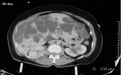
Normal drainage of bile may be impaired by cancer, benign strictures, or gallstones that obstruct the biliary pathways and  cause bile to accumulate above the obstruction. This dilates the ducts above the obstruction and forces bile to “leak” into the blood stream, turning the eyes, skin, and urine yellow – biliary jaundice. Biliary malignancy can occur in any segment of the bile ducts – within and without the liver – and frequently portends poor prognosis. Similarly, nonmalignant obstruction of the biliary tree can affect any segment of the tree, but it is not necessarily fatal.

Relieving malignant obstruction is important and can provide a patient quality time before death, if such is inevitable. This can be done surgically (by resecting the tumor and constructing a direct connection between the small bowel and the biliary tree, called choledochojejunal anastomosis), endoscopically (by deploying a stent across the malignant obstruction during endoscopic retrograde cholangiopacreatography (ERCP)), or percutaneously (by gaining access into the biliary tree through the skin and deploying a drainage catheter or a stent across the obstruction). The choice of the method of intervention depends on the patient’s preference, the skills available in an institution, and the location of the tumor; in general terms, non-resectable tumors should be approached endoscopically and stented or percutaneously if endoscopic intervention fails or is not feasible. The following cases of mine illustrate percutaneous biliary interventions.

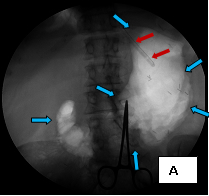
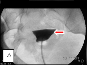
The first case (images A, B, and C), was an elderly man who came to me from a nursing home with advanced carcinoma of his common bile duct that severely narrowed the duct. The string of contrast outlining the very narrow common bile duct (red arrows, image A) and the dilated ducts above the narrowing (green and blue arrows, image A) testify to the severity of the disease. His left hepatic duct (blue arrow, image A) had been percutaneously decompressed elsewhere with a drainage catheter (yellow arrow, image A), but he remained jaundiced and ill.

Percut_biliary_decompression_1 Percut_biliary_decompression_2 Percut_biliary_decompression_3

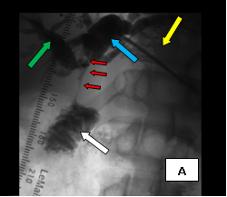
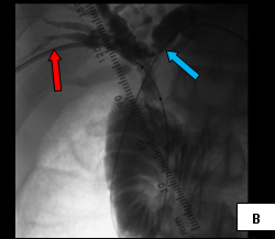
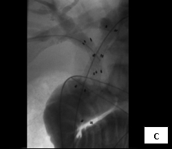
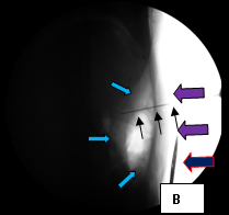
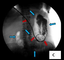
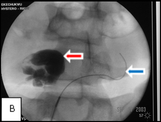
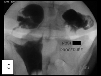
So, coming through a fresh percutaneous access on his right side (red arrow, image B) and through the drainage catheter already in his left hepatic duct (blue arrow, image B), I concurrently deployed stents across the obstructed confluence of the hepatic ducts and across the common bile duct. The effect of the treatment is evident by comparing images A and C: decompression of the dilated right and left hepatic ducts, relief of the critical obstruction of the common bile duct, and quick movement of copious amounts of the injected dye (which correlated with the movement of bile) into the duodenum.

Percutaneous_biliary_decompression_D Percutaneous_biliary_decompression_E Percutaneous_biliary_decompression_F

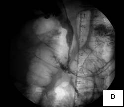
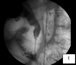
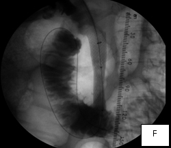
The second case was a woman with obstructive jaundice from cancer of the head of the pancreas, which produced a tapering narrowing of her common bile duct. In her, I crossed the diseased duct by cannulating a right intrahepatic duct from a right percutaneous puncture. Once I secured the access with a sheath, I was able to cross the obstruction with a stiff glidewire (image D) over which I deployed a Wallstent (image F).

Percutaneous_biliary_decompression_G

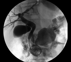
The third case illustrates percutaneous biliary drainage, which is the insertion of a drainage catheter from the skin, through the liver, into the duodenum, crossing the site of biliary obstruction. It is often the first step towards percutaneous biliary stenting and serves to decompress dilated biliary ducts and allow their inflamed walls to simmer down while waiting for the pathologic diagnosis of the obstructive disease before deploying a metallic stent across it. (Such staged approach is unnecessary if a non-metallic stent is employed in treating the disease, because such stents are easily retrievable should the final diagnosis recommend alternative forms of treatment; metallic stents, once deployed, are not removable.) Secondly, percutaneously stenting a biliary obstruction at the time of initial crossing is sometimes prolonged and challenging and risks seeding the blood stream with bacteria from the inflamed and friable biliary wall, causing sepsis.

In this woman, crossing her obstructed ampulla of Vater at ERCP failed, so I had to percutaneously deploy a biliary drainage catheter across it while waiting for the result of her biliary brushings. The single image illustrates the deployed catheter across the obstruction. 


Case 3: Percutaneous gastrostomy. Ken U. Ekechukwu, MD, MPH, FACP.

Sometimes feeding a patient by mouth is impossible or contraindicated, and providing them adequate nutrition becomes a challenge. Substituting oral nutrition with parenteral (intravenous) nutrition is fine when temporary, but not when prolonged; it has its own challenges and complications.

In such a situation, a patient can receive a gastrostomy or gastrojejunostomy feeding tube. Providing such feeding accesses involves the insertion of a feeding tube into the stomach (gastrostomy) or into the upper small intestine, the jejunum, through the stomach (gastrojejunostomy). In both cases the catheter placement is through the skin (percutaneous).  The pre- and post-procedure care of these patients is simple and the tube may be put to use within 24 hours, barring complications.

Percut_gastrostomy_1 Percut_gastrostomy_2 Percut_gastrostomy_3

These three images illustrate a variation of how I perform percutaneous gastrostomy.  Image A (supine frontal fluoroscope of the upper anterior abdomen) shows gastric insufflation after temporary gastroparesis with intravenous glucagon – distention of the stomach with air (the blue arrows outline the distended stomach) – through a nasogastric tube inserted into the stomach through the nose (the red arrows). The purpose of distending the stomach with air is to bring the anterior wall of the stomach against the anterior abdominal wall, which facilitates passing instruments of the procedure from the outside into the stomach. This is shown in Image B (a cross-table lateral fluoroscope of the upper abdomen) in which the light blue arrows outline part of the air-filled stomach; the purple arrows mark out the skin of the abdomen on which lies a pair of scissors, identifying the skin (the deep blue arrow with red outline); the black thin arrows indicate a 16 gauge needle passing from the surface of the skin into the lumen of the stomach. Image C (also a supine frontal fluoroscope of the abdomen) shows the deployed feeding tube (red arrows) within the stomach (light blue arrows). 

Percutaneous gastrostomy is quick and safe, typically requiring conscious sedation and the use of local anesthetic to do.  Proper care of the access requires adequate flushing of the tube after each use, preferably with fizzy drinks (carbonated beverages), good grinding of pills before administering them through the tube to prevent blockage of the tube, and meticulous attention to the skin around the opening into the catheter track to prevent infection.

Percutaneous insertion of a nephrostomy catheter. Ken U. Ekechukwu, MD, MPH, FACP.

Percutaneous nephrostomy is the insertion of a drainage catheter through the skin into the collecting system of a kidney to relieve obstruction of its drainage system.

Kidney cells produce urine which passes into the renal tubules and empties into the renal calyces. From the calyces, urine passes into the renal pelvis through the renal infundibula and from the pelvis empties into the urinary bladder through the ureter; it is then voided through the urethra.

Diseases of the urethra, the bladder, and the ureter may obstruct the flow of urine causing it to accumulate in and dilate the structures above the obstruction. Stones and strictures are the commonest causes of such obstruction.  Untreated and, depending on its severity, the pressure built up by the obstruction may damage the kidney cells or the static urine may become infected.

The obstruction may be relieved by the insertion of a ureteral stent through the urethra or, if this is not possible, the urine drained by the insertion of a tube into the renal pelvis through the skin (percutaneous nephrostomy). Such diversion of urine may be temporary or permanent depending on the nature of the obstructing disease and the feasibility of relieving the obstruction with a ureteral stent. 

The image below shows the final image after I carried out bilateral percutaneous nephrostomies in a man with bladder outlet obstruction due to prostate cancer.

Percutaneous_nephrostomy_catheter_insertions

Key to the image: 1 = Right renal calyx; 2 = Right renal pelvis; 3 = Right ureter; 4 = Urinary bladder; 5 = Pigtail catheter in right renal pelvis; 6 = Narrow prostatic urethra due to prostate cancer; 7 = Pigtail catheter in the left renal pelvis. Observe that the obstruction of the left structures is less severe than the obstruction on the right. The patient was prone for the procedure, hence the seeming switching of his sides.


Transcervical recanalization of obstructed fallopian tubes. Ken U. Ekechukwu, MD, MPH, FACP

One of the causes of infertility in women is obstruction of the fallopian tubes, the two trumpet-like canals that extend from the corners of the uterine fundus towards the ovaries. They conduct eggs released by the ovaries towards the inner chamber of the uterus called the endometrial cavity and may become occluded by disease, typically sexually transmitted disease. More commonly, however, obstruction of the fallopian tube at its cornu (the attachment of the tube to the uterus) is caused by refluxed menstrual debris, not pelvic inflammatory disease.  Tubal occlusion from whatever cause impedes the arrival of eggs from the ovaries to the endometrial cavity causing infertility.

The location and type of tubal obstruction vary: obstructions can occur close to or away from the uterine cavity and may be partial or total. Interventional radiologists can reopen fallopian tubal obstructions through a woman’s cervix by a procedure called transcervical recanalization of the fallopian tube. The closer fallopian obstruction is to the uterine cavity, the more likely transcervical recanalization will be successful; more distal obstructions are less reparable by this technique.

Transcervical_fallopian_tube_recanalization_1 Transcervical_fallopiantube_recanalization_2 Transcervical_fallopiantube_recanalization_3

Images A, B, and C belong to a young woman who was referred to me for hysterosalpingography because of infertility and was shown at the examination to have bilateral cornual occlusion of the fallopian tubes. Image A shows contrast opacifying her endometrial cavity (red arrow), but not the fallopian tubes due to bilateral cornual occlusions. In image B, I have reopened the right cornual occlusion, filling a large right hydrosalpinx with radiocontrast (red arrow), and have a guide wire in the left fallopian tube (blue arrow), which implies relief of the obstruction. Image C, the final image, shows that both cornual occlusions are relieved, but there are bilateral large hydrosalpinges because of fimbrial adhesions, proven by the absence of contrast spill into the peritoneal cavity.


Autosomal dominant polycystic kidney disease
Ken U. Ekechukwu, MD, MPH, FACP.

History
This is a 57-year old woman taken to a community hospital for care following a motor vehicle collision.

Radiologic findings

motor_vehicle_collision_CTAP_1motor_vehicle_collision_CTAP_2
Figure1: These are two contiguous computerized axial tomographic sections of the abdomen obtained after intravenous infusion of contrast.They show an enlarged liver that contains multiple cysts ranging in size from a few to several centimeters, diffusely spread throughout the organ. The kidneys also contain several (>5 total, bilaterally) similar though much smaller cysts but they are not very large. The pancreas, at least to the naked eye, is spared. There are no other obvious abnormalities; the lady, fortunately, had no significant intra-abdominal injuries from her trauma.

Radiologic diagnosis:  Autosomal Dominant Polycystic Disease (ADPD).

Radiologic differential diagnosis
Polycystic Liver disease.
Tuberous sclerosis.
Autosomal recessive polycystic disease (ARPD).
Von Hippel Lindau disease.
Multiple simple cysts in the liver and kidneys.

Discussion

The definitive diagnosis of this case hinges upon the patient’s family history, genetic evaluation and, if necessary, tissue procurement. Based on the radiographic appearance of her liver and kidneys, however, the two prime suspects should be ADPD and Polycystic liver disease. Of the two, the former is more likely because of commonality and the absence of hepatic fibrosis.

Hepatic cysts may be congenital or acquired. The acquired variety is due to trauma, inflammation, parasitic infection or tumor. The cysts are usually not as numerous as in this case. The congenital variants are rare and autosomal dominant; 50% of them may have polycystic kidney disease (1).

Tuberous sclerosis and von Hippel Lindau (vHL) disease belong to the phakomatoses, a group of neurocutaneous disorders that may have cysts in the viscera. The former may show bilateral asymmetric renal polycystic lesions but differs from ADPD by the presence of renal angiomyolipomas and involvement of the lungs, skin and brain.  vHL disease on the other hand comprises cerebellar hemangioblastoma, retinal hemangiomas and, occasionally, pheochromocytomas. This patient had none of these.

It is uncommon to have this degree of hepatomegaly and bilateral multiple renal cysts due to the presence of multiple simple cysts in the liver or the kidneys. The term ‘polycystic kidney disease’ encompasses both the autosomal dominant (ADPD) and the autosomal recessive (ARPD) forms of polycystic kidney disease. Although there is considerable effort to distinguish the two forms of the disease, for obvious genetic reasons, there are cases in the young in which the imaging features of the two forms of the disease are present in the same child, making it impossible to confidently allocate the disorder to a particular category. There have been reports of cases previously ascribed to one form of the disease by prenatal US that over time evolved to the other variant. Therefore, in children with polycystic kidneys imaging cannot hope to provide a conclusive diagnosis, but can complement clinical findings. In adults, the features of ADPD, a confirmed family history of the disease, and its imaging features make its diagnosis relatively easy.

In ARPD, there is always involvement of the liver by fibrosis with or without ectasia of the bile ducts similar to that seen in Caroli’s disease (6). The patient, always the index case in a family, typically presents younger but may appear in adolescence with hematemesis due to portal hypertension; the disease, though, is not compatible with longevity and is not a likely diagnosis in this case.

Autosomal dominant polycystic kidney disease (Adult polycystic kidney disease, Potter type III disease) is the fourth commonest cause of end-stage renal disease (5). Its inheritance is by autosomal transmission and the disease has 100% generational penetrance but a variable expressivity. At least there are descriptions of three genetic variants of the disease (4); 85% of cases (PKD1) are due to an abnormality at chromosome 16q13.3, while 15% (PKD2) are due to an abnormality at chromosome 4q21-23. The third genetic locus, yet unmapped, is a rare cause of the disease. In the disease, there is heightened predisposition to the formation of epithelium-lined cysts in renal tubules and Bowman’s capsules that grow to variable sizes surrounded by fibrotic tissue and that ultimately destroy the kidneys (4). There may be cysts in the liver (25-50%), pancreas (9%), kidneys, spleen, thyroid, ovaries, uterus, testis, seminal vesicles, epididymis and the urinary bladder. Other associated abnormalities include saccular (berry) aneurysms of the circle of Willis (10 to 16% of autopsy cases and as many as 41% of patients undergoing cerebral angiography), aortic root abnormalities, mitral valve prolapse, hypertension, azotemia, proteinuria, lumbar and abdominal pain and abdominal and inguinal hernias. Renal failure is usually a late manifestation.

Ultrasound is a useful screening tool for ADPD in adolescents and contrast-enhanced CT or MRI are recommended when the US results are equivocal or if there is need to exclude complications of the disease (e.g. renal cell carcinoma) or other differential diagnosis (e.g. Tuberorus sclerosis) (7,8,9). The complications of the disease include hematuria (due to a ruptured cyst), renal calculi (composed of urate or calcium oxalate), renal cell carcinoma (difficult to diagnose but not commoner in the ADPD population than in the general population) and chronic renal failure (which develops in 50% of patients) (3). The prognosis of the disease depends upon the development of renal insufficiency. When the serum creatinine level is consistently above 1.5mg/dL, the disease has entered a progressive phase of renal insufficiency. Risk factors that accelerate the progress of renal failure include: male gender, multiple pregnancies, the PKD1 genotype, hematuria, hypertension and proteinuria (3). Treatment is by hemodialysis, peritoneal dialysis or renal transplantation. ADPD patients on hemodialysis or peritoneal dialysis fair as well or better than patients with end-stage renal disease from other renal pathologies.

References
1. Zheng D, Wolfe M, Cowley BD Jr, Wallace DP, Yamaguchi T, Grantham JJ. Urinary excretion of monocyte chemoattractant protein-1 in autosomal dominant polycystic kidney disease. J Am Soc Nephrol. 2003 Oct;14(10):2588-95. 
2. Bisceglia M, Galliani CA, Senger C, Stallone C, Sessa A. Renal cystic diseases: a review. Adv Anat Pathol. 2006 Jan;13(1):26-56. Review.
3. Grantham JJ. Mechanisms of progression in autosomal dominant polycystic kidney disease. Kidney Int Suppl. 1997 Dec;63:S93-7. Review.
4. Grantham JJ.The etiology, pathogenesis, and treatment of autosomal dominant polycystic kidney disease: recent advances.Am J Kidney Dis. 1996 Dec;28(6):788-803. Review.
5. Fall PJ, Prisant LM.Polycystic kidney disease. J Clin Hypertens (Greenwich). 2005 Oct;7(10):617-9, 625.
6. Sgro M, Rossetti S, Barozzino T, Toi A, Langer J, Harris PC, Harvey E, Chitayat D. Caroli’s disease: prenatal diagnosis, postnatal outcome and genetic analysis. Ultrasound Obstet Gynecol. 2004 Jan;23(1):73-6.
7. Dimitrakov DY, Dimitrakov JD, Despotov TB. Ten-year clinical and ultrasonographic follow-up of siblings from families with autosomal dominant polycystic kidney disease. Folia Med (Plovdiv). 2002;44(4):10-2.
8. Lackner E, Jobges M, Schirmer F, Hummelsheim H.     Intracranial aneurysms and autosomal dominant polycystic kidney disease] Fortschr Neurol Psychiatr. 2002 Aug;70(8):438-42. German.
9. O’Neill WC, Robbin ML, Bae KT, Grantham JJ, Chapman AB, Guay-Woodford LM, Torres VE, King BF, Wetzel LH, Thompson PA, Miller JP.    Sonographic assessment of the severity and progression of autosomal dominant polycystic kidney disease: the Consortium of Renal Imaging Studies in Polycystic Kidney Disease (CRISP).  Am J Kidney Dis. 2005 Dec;46(6):1058-64

Acknowledgement
The author acknowledges the tireless work of Obidike Nwakudu, MD in the publication of this article. Dr. Nwakudu was a resident in the Division of Family Medicine at Mount Sinai hospital, Chicago when he did the work.