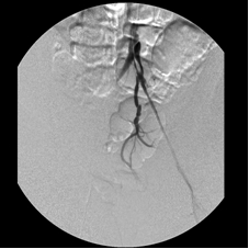
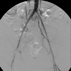
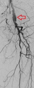
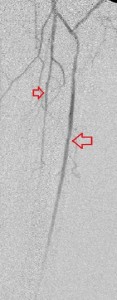
Author Archives: Ken Ekechukwu

Disabling left foot pain from left SFA disease | Ken Ekechukwu, MD, MPH, FACP

LM 2419105_prox_thigh_pre_reversed_imageLM2419105_prox_thigh_beforeLM2419105_prox_thigh_post
LM2419105_distal_thigh_beforeLM2419105_distal_thigh_post 

These images belong to a 48-year-old woman with insulin-requiring type 2 diabetes and long history of heavy cigarette smoking, who was referred to my interventional radiology clinic because of new pain in her left foot that substantially limited her life style, associated with color change. Her diabetes was uncontrolled and the pain seemed neuropathic in character, but the arterial doppler ultrasound of her lower limbs (not shown) uncovered insufficient perfusion of her left lower extremity. This was confirmed with computerized tomographic angiography (CTA) of her abdominal aorta with lower extremity runoffs to be due to chronic occlusive disease of her left superficial femoral artery. All the other examined vessels including her leg and pedal arteries were normal.

Faced with 3 possible reasons for her excruciating left foot pain – uncontrolled diabetes, arterial insuffuciency, and smoking- I decided to improve her left foot perfusion and her serum glycemic values in addition to assisting her quit cigarette smoking. Puncturing the opposite common femoral artery, I crossed over the aortic crotch to the left external iliac artery and used several devices including a stent-graft, to reopen her diseased left SFA. Her left foot pain and color change quickly resolved and at her last clinic visit for follow up she wore high-heel shoes – a thing she could not do before the intervention. She received prescriptions for Aspirin 81 mg daily, Plavix 75 my daily (6 months) and more help to cease smoking.

The top-row images illustrate her left SFA before treatment, while the lower-row images show the reopened vessel.


Left subclavian-vertebral steal syndrome. | Ken Ekechukwu, MD, MPH, FACP

PSxxx7724_LSCAstenosis_beforestenting PSxxx7724_LSCAstenosis_beforestenting_reversedimage
 PSxxx7724_LSCAstenosis_duringstentingPSxxx7724 LSCAstenosis_afterstenting

Key: LVA, left vertebral artery; LSCA, left subclavian artery; LCCA, left common carotid artery; LIMA, left internal mammry artery

The vertebral artery is the first branch of the subclavian artery and supplies the hindbrain, while the internal mammary artery is its second branch and courses behind the anterior chest wall to anastomose with the ipsilateral inferior epigastric artery in the rectus sheath; it offers a nautural route for lower body perfusion in obstructive diseases of the thoracic aorta or is grafted to a coronary artery in coronary artery bypass interventions (left internal mammary artery). In most people the left subclavian artery arises from the posterior aortic arch, while the right subclavian artery arises from the brachiocephalic trunk, itself the first branch of the aortic arch. A patient may develop vertebrobasilar insufficiency due stenosis or occlusion of the brachiocephalic or subclavian artery, called vertebral-subclavian syndrome. Similarly, symptoms of coronary hypoperfusion may develop in those in whom their left internal mammary artery (LIMA) is grafted to a coronary artery for coronary bypass surgery.

 In vertebral-subclavian steal syndrome, because the increased demand for blood that attends active use of an upper extremity is not met due to critical stenosis or occlusion of the brachiocephalic trunk or the subclavian artery, blood is short-circuted from the ipsilateral vertebral artery reducing hindbrain perfusion. The patient may experience such hindbrain symptoms as dizziness, nausea, dyequilibrium and vomiting that improve by resting the limb. Similarly, in coronary-subclavian syndrome blood is shunted from the coronary artery to which the LIMA is grafted, causing coronary syndrome involving the short-changed myocardial territory that improves with resting the left upper limb.

The commonest cause of large-vessel obstruction in the upper extremities is atherosclerosis and the left subclavian artery is affected by stenotic/occlusive disease 8 to 10 times more than the right subclavian artery. In the past such disease was solely managed by surgery – direct reconstruction via endarterectomy or aortic arch bypass – but with improved endovarscular techniques,  it is increasingly addressed endovascularly. Although few studies have compared endovascular stent-supported subclavian revascularization with aortic bypass interventions, the initial clinical success of the former is seen in nearly 100% of stenotic lesions and 60% to 100% of occlusions. Primary patency at 1 year has been reported in 91% to 100%, dropping to 82% to 86% at 3 years and 77% at 5 years.

 The 4 images you see above were recorded by me during primary stenting of irregular atherosclerotic stenosis of the left subclavian artery in a 54-year-old hispanic female who was referred to my interventional radiology clinic for management of the stenosis. She had presented to her primary care physician with symptoms of hindbrain ischemia upon using her left upper limb and earlier workup confirmed left subclavian arterial stenosis. The 2 top images illustrate the character and severity of her disease, the first image in the bottom row shows the process of balloon-expandable stenting of the disease, while the 2nd image in the bottom row reveals complete elimination of the stenosis and reappearance of the left vertebral and internal mammary arteires, which were invisible on the pre-intervention angiograms reflecting the severity of the stenosis.

Thrombosis of a left femoropopliteal bypass. | Ken Ekechukwu, MD, MPH, FACP

SHxxx4632_prox ltfempopbypass_before SHxxx4632_prox ltfempopbypass_after SHxxx4632_prox left leg_before SHxxx4632_prox left leg_after SHxxx4632_mid lfempopbypass_before
SHxxx4632_mid lfempopbypass_after SHxxx4632_distal ltfempopbypass_before SHxxx4632_distal ltfempopbypass_after SHxxx4632_distal left leg_before SHxxx4632_distal left leg_after

Key to images:

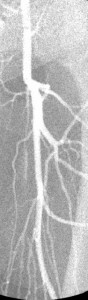
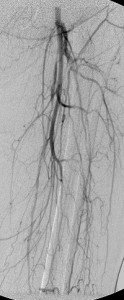
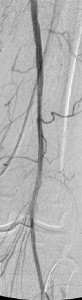
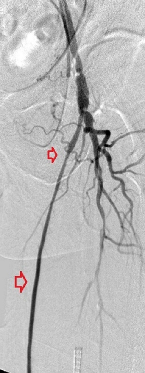
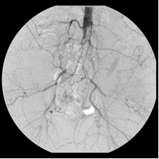
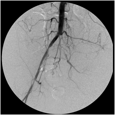
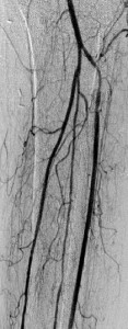
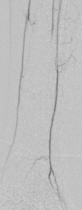
Top panel: Pre-intervention run-off angiogram of the left lower extremity showing, from left to right, irregular left common femoral artery (LCFA) arrowed on the 1st image, absent left superfical femoral artery (LSFA) or any bypass conduit on the 2nd image, sketchy descending collaterals from the left deep femoral (LDFA) that reconstitute a faint shadow of the left popliteal artery, arrowed on the 3rd image. The last 2 images faintly show three-vessel run-off below the left knee. The anterior tibial artery is most opacified, followed by the posterior tibial artery; the peroneal artery peeps through the upper edge of the last image. Note how weakly visible these vessels are due to the poor inflow from above.

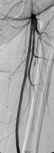
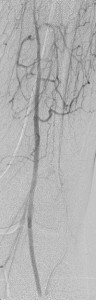
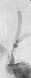
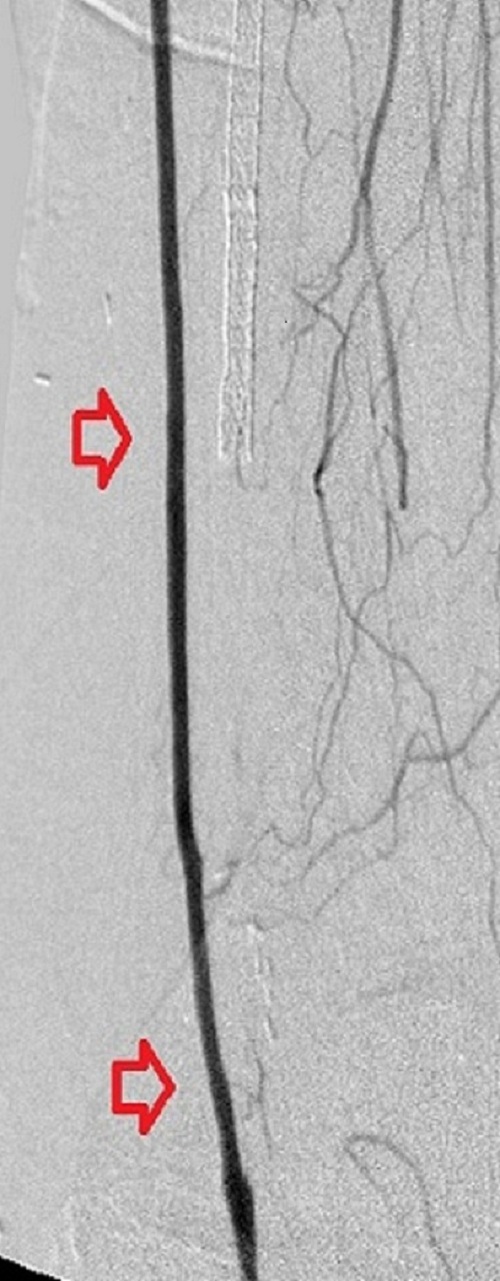
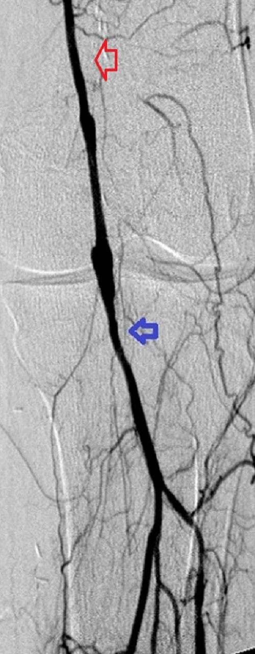
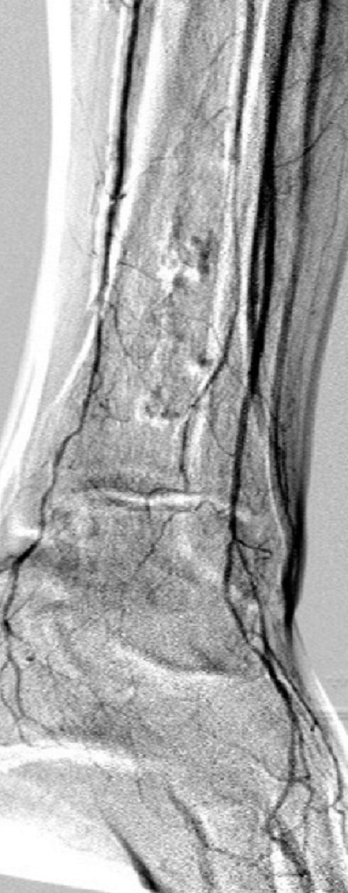
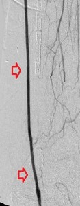
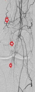
Bottom panel: Post intervention run-off arteriogram of the left lower extremity showing, from left to right, the proximal and distal segments of the re-opened left femoro-popliteal bypass (red arrows on images 1, 2, and 3). Contrast the full-column opacification of the below-knee left popliteal artery, arrowed blue on the 3rd image, and the enhanced visibility of the three-vessel subpopliteal domain to their vestigial appearances on the pre-intervention images, when they were poorly fed through collaterals.

Arterial bypasses are surgical contructs that connect normal proximal and distal segments of an arterial tree to deliver more blood  distally by avoiding a diseased segment of the tree. The preferred bypass conduit is a patient’s native vein, but when such is unavailable or what is available is unacceptable or insufficient, a synthetic conduit is used. Bypasses may be long or short depending on the length of the arterial obstruction. Their longevity varies and depends on the nature of the underlying arterial disease, the flow of blood into and out of them, the technical challenge in creating them, the skill of the operating surgeon, a patient’s blood pressure, and the nature of the bypass conduit. 

Bypasses fail if blood flow through them is restricted by upstream arterial disease or short-circuited through large proximal side trunks; by a poor distal run-off, a diseased pedal arch or a weak, impoverished, or discontinuos link between the two; by hypotension, especially soon after their creation, because this induces slow flow through them and hastens clot formation; synthetic conduits tend to stay open shorter than native veins. Finally, technically-challenging creations or those constructed by inexperienced hands tend to fail quicker than their counterparts.

Early bypass failure, within 30 days, precipitates acute ischemic symptoms and demands urgent surgical intervention. It frequenctly is due to unattended disease in the inflow or outflow channels, a mechanical error in its creation, hypotension, or problem with the conduit. Later failure causes less dramatic symptoms that demand less speedy intervention and, when it happens within 1 to 2 years of a bypass’ creation is often due to the development of anastomotic intimal hyperplasia. Intimal hyperplasia is physiologic smooth muscle enlargement in the wall of a native vein when it is subjected to high-pressure arterial flow and can be treated with balloon angioplasty. Bypass failure after 2 years is frequently due to progression of the underlying atherosclerosis, either in the inflow or the outflow vessels. There are times, however, when no identifiable cause for a failure exists and this is more prevalent with synthetic conduits.

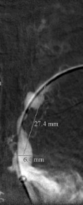
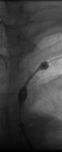
When blood flow into or out of a bypass fails, it thromboses and its salvage includes gaining access into it and removing the clot in addition to determining the reason for its failure. Such clot removal may be mechanical, as is frequently the case in acute graft failure, or through thrombolysis, as is the case in later failures. The above images illustrate the later scenario in which the patient presented about 1 year after a left femoropopliteal bypass was fashioned for them. I crossed into the lumen of the bypass conduit from a right common femoral arterial puncture and advanced an infusion cather into it for overnight continuous alteplace infusion following a bolus dose. (I favor 5 to 10 mg of alteplace bolus, followed by continuos infusion at 0.5 mg per hour, in company with fixed unfractionated heparin infusion at 500 units to 600 units per hour after a bolus dose of 3000 units to 5000 units). In this case the bypass proved to be a vein conduit connecting the left common femoral artery, proximally, to the mid popliteal artery, distally, without intimal hyperplasia. The cause of the failure was diminished inflow due to left iliac disease.The final runoff images reveal a three-vessel tibial domain continuous with a near-normal plantar arch.

Endovascular restoration of blood flow across total occlusions of the distal aorta and bilateral iliac channels. Ken U. Ekechukwu, MD, MPH, FACP.

This 41-year-old woman smoked for a long time and developed severe bilateral calf cramping after short walks. Investigation revealed that she had completely occluded her distal abdominal aorta and her right and left iliac arteries, severely restricting the flow of blood into her lower limbs. She was unwilling to have surgery and came to me for minimally invasive treatment. By dripping urokinase (a clot dissolver) over 72 hours through two catheters that I advanced across the occlusions and later stenting the opened arteries, I wholly restored blood flow through her iliac channels. She walked again – without cramps.

Iliac_arteries1 Iliac_arteries2 Iliac_arteries3 Iliac_arteries4