Author Archives: Ken Ekechukwu

Neurocysticercosis.
Ken U. Ekechukwu, MD, MPH, FACP.

History
This is an eighteen-month old child born of Mexican extraction resident in Texas, USA . His prenatal and natal histories are unremarkable.

Sagittal_T1_WI_A Sagittal_T1_WI_B
Sagittal_T1_WI_C Sagittal_T1_WI_D

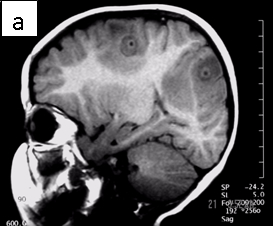
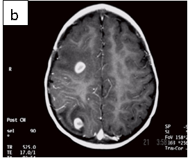
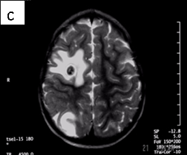
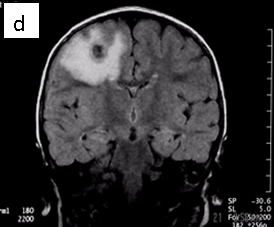
Figure 1: Neurocysticercosis in an 18-month child. a. Sagittal T1 without gadolinium; b. Axial T1 with gadolinium; c. Axial T2; d. Coronal FLAIR

Radiologic diagnosis: Neurocysticercosis.

Radiologic differential diagnosis
1. Pyogenic abscesses.
2. Tuberculomas.
3. Metastases.
4. Tuberous sclerosis.
5. Toxoplasmosis.

Discussion
Neurocysticercosis results when encysted larval forms of Taenia solium (pork tapeworm) invade the central nervous system (2). It is the most common parasitic disorder of the CNS (5) occurring in 4% of autopsy series from endemic countries of the world (in Latin America, Asia, Africa and some European countries).

Table 1: Cestode infections in humans 
 SPECIES  STAGE SEEN IN MAN  COMMON NAME  DISEASE IN MAN
 Taenia saginita  Adult  Beef tape worm  Rarely symptomatic
 Taenia solium  AdultLarva  Pork tape wormCysticercosis  Rare symptomaticCysts in the brain and other tissues
 Taenia multiceps  Larva  Bladder worm, coenurosis Cysts in the brain and eyes
 Echinococcus granulosus  Larva  Hydatid cyst disease  Solitary tissue cysts
 Echinococcus multilocularis  Larva  Alveolar cyst disease  Multilocular cysts
 Diphyllobotrium latum  Adult  Fish tape worm  Pernicious anemia
 Hymenolepis nana  Adult  Dwarf tape worm  Rarely symptomatic
Spirometra mansonoides Larva Sparganosis Subcutaneous larvae

Taenia solium is one of the eight cestode species that infect man (see Table 1). Humans are the definitive host for the parasite (we harbor the adult worm) while omnivorous or herbivorous vertebrates (like pig) host its larval form (intermediate host). Although the adult forms of the cestodes that we host rarely cause us harm, humans suffer variable degrees of illnesses when they host the larval forms; in the former case the parasites often reside harmlessly in the bowels, while in the latter they invade the tissues. For example, in its normal life cycle, humans harbor the pork tapeworm and shed its eggs (either individually or in proglottides impregnated with eggs) in our feces. The pig, an omnivore and its natural intermediate host, consumes the eggs of the parasite, which hatch into larval forms in its alimentary tract and invade its tissues. Consumption of undercooked meat from infected pork by us completes the cycle, freeing the encysted larvae when they encounter our gastric acid and bile salts. In contrast, we act as the intermediate host of the parasite when we ingest the tapeworm eggs by eating food infected with them (the infant’s mother in this case may have infected her child this way). Their larvae invade the bowel walls and invade our tissues as far afield as the brain, eyes, muscles, heart and other places wreaking havoc in their wake (7). In the CNS, the cysticerci (the larvae) may lodge in the brain parenchyma, the spinal cord, the subarachnoid space or the ventricles, lying dormant for years or causing various categories of clinical disease (see Table 2) (3).

Table 2: Clinical categories of neurocysticercosis
SYNDROME CLINICAL FEATURES
Acute invasive stage Commoner in children; mortality, 10%. Occurs immediately after the infection with fevers, headaches, and myalgias. Heavy infection may result in coma with rapid deterioration (cysticercal encephalitis). Treat aggressively with anti-parasitic and anti-inflammatory agents.
Parenchymal CNS cysticercosis 50% of cases; there is established parenchymal disease with seizures, focal deficits, intellectual impairment, personality changes and signs of raised ICP when severe.
Subarachnoid cysticercosis 30% of cases; there is larval invasion of the subarachnoid space including the cisterns causing disturbed CSF flow, sensorial changes and, as in other chronic basal meningitis, signs of vasculitis and parenchymal infarctions.
Intraventricular cysticercosis 15% of cases; there are cysts in the ventricles, the 4th most frequently involved, causing intermittent obstruction to CSF flow with head movements. The aggressive form is racemose cysticercosis in which sheets of parasites spill out into the subarachnoid space severely impairing CSF drainage. It is commoner in young women
Spinal cysticercosis Cord compression with radiculopathy, transverse myelitis and signs of meningitis.
Ocular cysticercosis Eye pain with scotomata, iridocyclitis, clouding of the vitreous and retinal inflammation or detachment

However, 2 to 10 years after CNS invasion, the dormant cysts may die, lose osmoregulation, absorb fluid and disintegrate, releasing antigens that set up variable degrees of inflammation. The clinical conundrum that results from CNS larval invasion depends upon the size of the invasion, and the location and degree of the inflammation. You can make a sure diagnosis of neurocysticercosis by analyzing infected tissue microscopically. But you can make a presumptive diagnosis of the disease if your patient is from or resides in an endemic area (as in this case) and, if laboratory analysis of their CSF specimen including an immunoblot test, and their CNS imaging results are positive for markers of the disease (4,5) (see Table 3). 

 Table 3: Diagnosing neurocysticercosis with laboratory and radiologic information
CSF analysis Hypoglycorrhachia.
Elevated CSF protein.
Lymphocytosis and eosinophylic pleocytosis (5-500 cells/microliter).
ELISA and Western blot testing for specific IgM and IgG anticysticercal antibodies in the CSF (75 to 100% sensitivity).
CT scan A variable appearance including multiple, low-density lesions, 0.5 to 2.0cm in diameter. In acute disease, they enhance after contrast administration, surrounded by vasogenic edema with or without mass effects. Dead parasites (not dying parasites) show as non-enhancing calcified 5mm bodies.
MRI Live forms have a characteristic appearance: fluid-filled lesions containing an inverted scoleces, surrounded by thin low-signal capsule. They do not stimulate inflammation and do not enhance; dying forms do. In the less common racemose type, the cysts may be hard to see because they have similar imaging features as the CSF.

For cysts that cause symptoms outside the CNS, surgical resection achieves cure. The treatment of symptomatic neurocysticercosis, which carries a 50% mortality rate, is more problematic. Two drugs, albendazole and praziquantel control symptoms and cause regression in the size and number of cysts in patients with viable (non-enhancing) cysts in their brain parenchyma. However, they provide limited improvement in patients with arachnoiditis and none in patients with intraventricular cysts. These latter patients should be treated with surgery or palliated with ventricular shunting, anticonvulsants, and anti-inflammatory drugs. According to our case’s physician, he did not receive anti-parasitic medication because the imaging features suggested that the parasites were dying (vasogenic edema and ring enhancement) (6).

Two cautionary injunctions about treatment, though: first, 20% of patients with parenchymal cysticercosis worsen symptomatically following the institution of drug treatment as the parasites die and release their antigens. Concomitant administration of anti-inflammatory drugs subdues this phenomenon. Second, because anti-inflammatory agents alter the CNS pharmakokinetics of the anti-parasitic agents, their routine use is discouraged.

You should rescan your patients 3 months after therapy to judge their response to treatment; prescribe an alternate drug to the one used ab initio if there is no response.

For patients with ocular cysticercosis (remember, 20%), you are better off holding drug treatment until resection of their lesions because they do not respond well to medication.

Bibliography
1. Shandera WX, White AC Jr, Chen JC, Diaz P, Armstrong R. Neurocysticercosis in Houston, Texas. A report of 112 cases. Medicine (Baltimore). 1994 Jan;73(1):37-52.
2. Domenici R, Matteucci L, Meossi C, Stefani G, Frugoli G. Neurocysticercosis: a rare cause of convulsive crises Pediatr Med Chir. 1995 Nov-Dec;17(6):577-81. Italian.
3. Caparros-Lefebvre D, Lannuzel A, Alexis C, Strobel M, Janky E Cerebral cysticercosis: why it should be treated. Presse Med. 1997 Nov 1;26(33):1574-7. French.  
4. Ruiz-Garcia M, Gonzalez-Astiazaran A, Rueda-Franco F. Neurocysticercosis in children. Clinical experience in 122 patients. Childs Nerv Syst. 1997 Nov-Dec;13(11-12):608-12.
5. Grill J, Pillet P, Rakotomalala W, Andriantsimahavandy A, Esterre P, Boisier P, Guyon P. Neurocysticercosis: pediatric aspects Arch Pediatr. 1996 Apr;3(4):360-8. Review. French.
6. Riley T, White AC Jr. Management of neurocysticercosis. CNS Drugs. 2003;17(8):577-91. Review.
7. Rahalkar MD, Shetty DD, Kelkar AB, Kelkar AA, Kinare AS, Ambardekar ST The many faces of cysticercosis. Clin Radiol. 2000 Sep;55(9):668-74.

Acknowledgement
The author acknowledges the untiring work of Obidike Nwakudu, MD in the preparation of this article, while he was a resident in Family Medicine at Mount Sinai hospital, Chicago, IIinois.

Neurofibromatosis. Ken U. Ekechukwu, MD, MPH, FACP.

History
The patient is an elderly woman seen in a clinic because of persistent mid-back pain. She did not return to her primary care physician after this study; little of her history is available. These are the only imaging studies available in her records.  

Radiologic findings
The radiographs are frontal and lateral views of her thoracic spine.  They show:  
1. Diffuse osteopenia.
2. Levoscoliosis of the mid and lower thoracic spine.
3. Compression fractures of the eighth and ninth thoracic vertebrae.
4. Numerous round well-defined soft-tissue masses attached to the chest wall.

The key to the radiologic diagnosis of this case lies in the non-calcified cutaneous masses of varying sizes. It is easy to assume the masses are within the patient’s thorax but a closer look shows that most are well-defined, have lucent halos around them and, so, are  external to the chest wall. They are pathognomonic of von Recklinghausen’s disease (neurofibromatosis type1). The patient is osteoporotic and the vertebral compression fractures may be due to this.

Neurofibromatosis


Vertebral compression fractures are common in the elderly, especially in the setting of osteoporosis. They may also occur in conditions that weaken the vertebrae such as metastatic disease, infection, Scheuermann’s disease (multiple, contiguous intraosseous herniation of nucleus pulposus at the center of weakened endplates (multiple Schmorl’snodes)), sickle-cell anemia and histiocytosis X. Again, the cutaneous masses make the diagnosis easy.   Scoliosis and kyphoscoliosis of the spine may be congenital or acquired. When congenital they are due to failure of segmentation or formation of vertebral bodies; they may be mixed, showing features of both.   There are many acquired causes of scoliosis and kyphoscoliosis (see below) and painful conditions and von Recklinghausen disease are two such entities.  The upper and lower vertebrae in the patient show subtle signs of formation failure suggesting that in her the scoliosis is congenital (a component of the von Recklinghausen conundrum). Clearly, pain from the compression fractures may contribute to the abnormal spinal curvature. Thus, in the patient, congenital and acquired scoliosis may be at play.  

Radiologic differential diagnosis
For multiple cutaneous masses:
The cutaneous masses in this patient are pathognomonic for neurofibromatosis
For scoliosis:
1. Mesodermal and neuroectodermal diseases including neurofibromatosis, Marfan’s syndrome,   homocystinuria.
2. Neuromuscular diseases such as myelomeningocele, spinal muscular atrophy, Friedrich’s ataxia, poliomyelitis, cerebral palsy, and muscular dystrophy.
3.
Painful scoliosis as in osteoid osteoma, osteoblastoma, intraspinal tumor, and infection.
4. Congenital scoliosis due to failure of formation, failure of segmentation, or mixed defects.
For multiple collapsed vertebrae:
1. Osteoporosis
2. Neoplastic disease
3. Scheuermann disease
4. Infection
5. Histiocytosis X
6. Sickle-cell anemia

Discussion
Neurofibromatosis type 1 (NF1) is the most common of the phakomatoses, a heterogeneous group of histiogenetic disorders characterized by the presence of central nervous system and, for the most part, cutaneous manifestations. Many also have prominent visceral and connective tissue abnormalities.

Box 1: The Phakomatoses. 
Neurofibromatosis
    Neurofibromatosis type 1
    Neurofibromatosis type 2
    Segmental neurofibromatosis.
Von Hippel-Lindau syndrome
Sturge-Webber syndrome
Tuberous sclerosis
Others
    Ataxia-Telangiectasia
    Rendu-Osler-Weber
    Klippel-Trenaunay-Weber
    Neurocutaneous melanosis
    Wyburn-Masosn Basal Cell Nevus
    Cowden Disease.


NF-1 occurs in one of 2000 to 3000 births. It is inherited by autosomal dominant transmission but has a high spontaneous mutation rate (50%) so that it may occur in offsprings of unaffected parents. The penetrance of the disease is high but its expressivity variable. The problem in the disease lies in the long arm of chromosome 17; there is neuroectodermal and mesodermal tissue dysplasia with potential for diffuse systemic involvement. Males and females are affected equally.

 
The clinical diagnosis of the disease hinges upon the demonstration of the characteristic cutaneous manifestations or two of the set of criteria shown below. The case in question has numerous cutaneous masses that are consistent with disease although we do not have a full account of the patient’s history.
 
 The NIH Consensus Development Panel developed a set of diagnostic criteria for NF-1 that includes the following:
 1. 6 or more 5mm or larger café-au-lait spots.
 2. 1 plexiform neurofibroma or two or more neurofibromas of any type.
 3.  2 or more pigmented iris hamartomas (so-called Lisch nodules).
 4.  Axillary or inguinal region freckling.
 5.  Optic nerve glioma
 6.  First-degree relative with NF-1
 7.  Presence of a characteristic bone lesion (e.g. dysplasia of the greater wing of the sphenoid, pseudoarthrosis).
 
The disease may affect many organs including the brain, spinal cord, peripheral nerves, bones, the dura, the eye and the blood vessels (see below).

Box 2: Organs that may be affected by NF-1 
Non-CNS lesions
    Visceral and endocrine tumors:
    MEA IIb
    Musculoskeletal lesions:
    Ribbon ribs
    Pseudoarthroses
    Tibial bowing
    Focal overgrowth of digit, ray or limb
CNS lesions
    Optic glioma
    Nonoptic glioma
    Basal ganglionic and white matter hamartomas
    Spinal cord, roots/ peripheral nerves:
    Hamartomas
    Cord astrocytoma
    Neurofibromas
    Osseous and dural lesions:
    Hypoplasia of the greater wing of the sphenoid bone
    Sutural defects
    Kyphoscoliosis
    Meningoceles
    Ocular/orbital manifestations:
    Optic nerve glioma
    Lisch nodules
    Buphthalmos
    Retinal phakomas

NF-1 is different from NF-2 although it, too, is inherited by autosomal dominant transmission. NF-2 is less common than NF-1 (1:50,000), is due to defects in chromosome 22 and in it, cutaneous manifestations are rare. The dominant mode of presentation of NF-2 is by central nervous system lesions (approximately 100%). These include schwannomas of cranial and peripheral nerves, meningiomas in the brain and spine (often multiple), nonneoplastic intracranial calcifications (especially choroids plexus), spinal cord ependymomas and secondary changes of the spine.

 
References
1. Smirniotopoulos JG, Murphy FM: The phakomatoses, AJNR 13: 725-746, 1992.
2. Huson SM, Harper PS, Compston DA: von Recklinghausen neurofibromatosis: a clinical and population study in southeast Wales, Brain 111:1355-1381, 1988.
3. National Institute of Health  Consensus Development Conference: Neurofibromatosis Conference Statement, Arch Neurol 45: 579-588, 1988
4. Diagnostic Neuroradiology, Anne G. Osborn

Preparing patients for interventional radiological procedures. Ken U. Ekechukwu, MD, MPH, FACP.

Most interventional radiological (IR) procedures are safe and help patients immensely, but sometimes they are associated with complications. Some complications are inherent to the nature of a procedure, while others are not and are preventable by taking some precautions before, during, and after a procedure.  Here, I offer general guidelines to physicians and patients on how to prepare for IR procedures.

There are specific preparations for individual procedures. These are usually explained to the patient at the interventional radiology clinic or in their room (if they are hospitalized); such explanation should be part of the process of obtaining an informed consent for a procedure. You may review them at this web site under “About IR procedures and tests”, or, visit www.sirweb.org/patients for more information.  

Diet, medications, and hydration
Although many interventional radiological (IR) procedures are performed with only local anesthesia, a majority require conscious sedation for the patient’s comfort and alleviation of pain and anxiety. A few require more sophisticated anesthesia. Since the medications used to induce conscious sedation depress respiration and consciousness (and some directly stimulate the vomiting center in the brain stem), administering them to patients with full a stomach is risky since they may vomit and choke themselves during a procedure. It is important then that the stomach is empty when a patient receives conscious sedation. Generally, different meals clear the stomach at different rates, water and other liquids clearing the fastest, while fatty meals clear the slowest; carbohydrates and proteins lie in between, carbohydrates clearing quicker than proteins. Most meals will vacate the stomach by 8 hours. Herein lies the justification for having nothing by mouth after the midnight preceding the morning of a procedure, the assumption being that there would be ample time for the contents of the stomach consumed at midnight to vacate it before a procedure slated for 8:00 am or thereabout. Patients may, however, drink clear water until 2 hours before the start of a procedure; in fact they are encouraged to do so in some instances, when adequate hydration is necessary to prevent renal injury from iodinated radiocontrast.

Exposure to iodinated radiocontrasts (contrast agents) deserves special mention. Iodinated radiocontrasts are chemical agents rich in iodine that are given to patients either by mouth or intravenously to improve the visibility of normal and abnormal tissues in the body when x-rays pass through them. Since abnormal tissues have more blood vessels most of which abnormally ‘leak’ intravenously administered radiocontrast into the tissue interstices, delaying its washout, they stand out in recognition more than normal tissues, the so-called contrast enhancement. This is the reason intravenous contrast enhancement is encouraged in studying such disorders as tumors, infections, inflammations, radiation injuries, post-surgical changes, etc., – situations in which the examiner wishes to leverage or take advantage of the differences in the number and abnormalcy of blood vessels in diseased tissues to distinguish them from normal ones. Such use of radiocontrast is employed in IR in addition to its use in opacifying the lumens of blood vessels when working on them.

Iodinated radiocontrasts given by mouth, to a large degree, cause no harm, but those given intravenously can alter renal function in some patients. These include those older than 60 years; those with existing poor renal function; those who are dehydrated; those with multiple myeloma; and diabetics. People who belong to one or more of these groups are at increased risk of contrast-induced nephropathy, a state of diminished renal function following exposure to an iodinated radiocontrast. Their risk for this may be reduced by sufficient hydration with normal saline and the administration of such free-radical scavenger as acetyl cysteine (Mucomyst) before exposure to the radiocontrast. Of course, it is important to weigh the risks of such exposure against its potential merits before proceeding with a test or procedure; there are moments when doing nothing (masterly inactivity) is wiser and safer than taking an action. Adequate intravenous hydration of a patient before exposure to iodinated radiocontrast is second only to no exposure to it in the prevention of contrast-induced nephropathy.

Diabetic patients on metformin are a special case because the concurrent taking of the drug and the development of renal impairment can cause lactic acidosis. Lactic acid is one of the many products of metabolism excreted by the kidneys. When it exists in excess the condition is called lactic acidosis. Any form of renal impairment will reduce the excretion of metformin and lactic acid leading to type B (non-hypoxic) lactic acidosis. This condition, though rare, is reported to have a 50% mortality rate. Metformin is a biguanide oral hypoglycemic agent that works by increasing the movement of sugar into glucose-utilizing cells while suppressing the synthesis of sugar in the liver (gluconeogenesis). It is excreted by the kidneys and, like other biguanides, suppresses the action of pyruvate dehydrogenase as well as decrease the transport of mitochondrial reducing agents. These two actions accumulate pyruvic acid in the cell that is converted into lactic acid.  Since contrast-induced nephropathy is associated with diminished renal function, diabetic patients (who are already at increased risk for contrast-induced nephropathy) taking metformin should switch to non-biguanide oral hypoglycemics or insulin starting on the day of radiocontrast exposure until their blood chemistry taken 48 hours to 72 hours after the  exposure documents normal renal function.

Prophylactic measures
Some complications of interventional radiological (IR) procedures like bleeding at an arterial puncture site, systemic or local skin infection, or acute or delayed thrombosis of a revascularized artery are preventable.

The process of re-establishing blood flow through an occluded artery can be complicated and challenging. It also traumatizes the arterial wall and may precipitate acute re-thrombosis or occlusion of the vessel during or shortly after the procedure. Not only does this jeopardize the interventionalist’s work, it also re-exposes a patient to the ordeal of repeating the intervention at enhanced risk of complications. For these reasons, interventional radiologists give oral Aspirin and clopidogrel (Plavix) (both of which are called antiplatelets because they prevent platelet aggregation) to their patients before starting arterial interventions and intravenous heparin (and sometimes other antiplatelets drugs) during the procedure to reduce the risk of acute thrombosis; patients continue oral clopidogrel for 3 to 6 months afterwards and Aspirin for life.

The commonest reason for bleeding at an arterial puncture site is uncontrolled hypertension, of course assuming a normal coagulation profile. Therefore, hypertensive patients are encouraged to take their morning dose of antihypertensive (s) before arriving for their procedure.

Diabetics on insulin should skip the morning dose of their short-acting insulin and take half their dose of the long-acting insulin; they should receive D5/0.45 normal saline infusion for maintenance therapy and have their blood sugar monitored before, during, and after the procedure. Importantly, their procedure should be done as early in the day as is possible to avoid hypoglycemic attacks.

Infections are known to complicate procedures done on certain parts of the body: the skin, the alimentary tract, and the genitourinary tract. They can also complicate interventions that devitalize tissues like embolizations. Sometimes, mere knowledge of the hazards of infection complicating a procedure on certain anatomic areas of the body, such as vertebroplasty, informs the decision for antibiotic coverage. In all these circumstances it is prudent to provide patients with prophylactic antibiotic coverage and the choice of the antibiotic is governed by the prevailing body flora of the area of the procedure and local microbiologic idiosyncrasies. There are now guidelines for this provided by the Centers for Medicare and Medicaid Services (CMS) in association with other recognized associations, and their use is mandatory.  The reader is encouraged to visit www.cms.gov for more information on this.

Benign ovarian cystic teratoma. Ken U. Ekechukwu, MD, MPH, FACP.

History
This is a 20-year old female who visited the emergency department because of pain in the left lower quadrant of her abdomen. She had no significant past medical problems, but on examination had an excessive amount of hair on her lips, chest, and lower belly. Her pubic hair, though also excessive, was feminine in its distribution. While in the emergency department she had contrast-enhanced CT scan of her abdomen and pelvis, but had no other imaging studies. Measurements of her testosterone while on admission were normal. Later, she underwent open laparotomy and had her abdominal mass removed.

Radiologic findings

benign_cystic_ovarian_teratoma1

This is a single axial section through the patient’s false pelvis showing a midline, thin-walled (1mm), complex  mass. The mass measures 12cm x 13cm x 13cm and at its 10 to 12 o’clock position is a heterogeneous mass of tissue (Rokitansky protuberance, dermoid plug) comprising a high-density rectangular structure (coarse calcification, mal-formed tooth), and lobules of tissue isodense to subcutaneous fat (fat). The preponderance of the mass, however, is hypodense to enhanced muscle but slightly hyperdense to noncontrast urine (not shown); it is fatty debris. The mass is anterior to the rectum, straddles the pelvis and displaces the small bowel to the sides. The ovaries are not discernible. The appearance of the mass is pathognomonic for a mature teratoma.

Differential radiologic diagnosis
There is no differential diagnosis. The radiologic features of the lesion are pathognomonic for benign cystic teratoma of the ovary.

Gross pathologic findings
A large mass which on sectioning revealed:
1. Cheesy liquid debris.
2. A solitary tooth.
3. Thick golden-yellow congealed fatty tissue.
4. A loch of hair.
Please see below.

benign_cystic_ovarian_teratoma2

Final pathologic diagnosis
Mature (Benign) Cystic Teratoma.

Discussion
Teratomas (teratos, Greek for monster) are congenital tumors of derivatives of all three germ layers. They arise from pluripotent embryonal cells and occur in the ovaries, testes, mediastinum, retroperitoneum, and the sacrococcygeal region, in order of decreasing frequency. The location of the tumor in our case suggests that it is ovarian in origin; presacral or retroperitoneal origins are less likely alternatives.

Teratomas are the most common tumors of the ovary in children and adolescents although they may present at any age in life. They may be mature (cystic) or immature (solid, a.k.a embryonal) and carry a 0.25-1.5% risk of malignant degeneration, representing 1% of malignant ovarian tumors. The risk of malignant degeneration is predicted by the presence of neuroectodermal remnants, which the present case lacks.

Two-thirds (67%) of the cases present as an abdominal mass (as in this case) and 10% – 15% are bilateral. Other modes of presentation include abdominal pain (our case), ovarian torsion, nausea, vomiting and, rarely, ascites. There may be rare association with hyperprolactinemia, hemolytic anemia, hyperthyroidism (struma ovarii) or Mayer-Rokitansky-Kuster-Hauser syndrome. The latter consists of a lack of mullerian development, congenital absence of the vagina and rudimentary uterus (typically bicornuate remnants), with normal uterine tubes, ovaries, secondary female sex characteristics and normal growth.  There was mention to me by one of the patient’s physicians that she had a small uterus and was hirsute. I doubt that we can invoke Mayer-Rokitansky-Kuster-Hauser syndrome in this case because of the absence of the other features and I doubt that her alleged hirsutism has any relationship to the teratoma. She recuperated well from her surgery and went home in good condition.

References
1. Friedman AC, Pyatt RS, Hartman DS et al 1982. CT of benign cystic teratomas. AJR 138: 659-665.
2. Buy JN et al. Cystic Teratoma of the ovary: CT detection; Radiology 171: 699-701.  1989
3. Kallenberg GA et al. Ectopic hyperprolactinemia resulting from an ovarian teratoma: JAMA, 263: 2472-2474.

Stenting of the carotid artery. Ken U. Ekechukwu, MD, MPH, FACP.

What is stenting of the carotid artery?
The carotid artery carries fresh blood from the heart to the neck, the face, and the head including the anterior brain. Two carotid arteries arise from the aorta, the large artery in the chest that comes directly from the left heart; one runs through the right neck, while the other runs through the left. Somewhere close to the angle of the jaw the arteries divide into two unequal branches that carry blood to the front of the brain (the larger artery) and the neck and face (the smaller branch), respectively.

These arteries, like all arteries in the body, can be narrowed or obstructed by cholesterol-rich material (called atheromatous plaque or, simply, a plaque) that grows in their walls due to conditions like hypertension, diabetes, high cholesterol, and smoking. This narrowing, when severe or complete, slows or shuts off blood flow to the brain causing stoke-like symptoms or actual stroke. Sometimes tiny particles of the plaque break off and travel with blood into the brain to cause stroke. Stroke can be prevented from occurring in some patients by doing surgery on the narrowed artery (Endartherectomy) to repair it or by widening it with a stent. A stent is cylindrical tube made from metal into whose walls are cut numerous small holes that turn the walls into a mesh. A stent placed across a narrowed or blocked artery keeps it open and reduces the chance of particles of atheroma breaking off to cause a stroke.

How does one prepare for stenting of the carotid artery?
You must prepare yourself emotionally for the procedure and gather as much information as you can. Your health care provider will tell you what the procedure entails and discus its benefits and risks with you. Expect to be asked to take Aspirin and clopidogrel (Plavix) for 3 to 5 days (if you are not already taking them) before the procedure. After the procedure you will take Aspirin for life and Plavix for 3 to 6 months. These medications reduce the chance of clot formation in the arteries or the stent during and after the procedure. You will not be placed under general anesthesia because staying awake during the procedure allows your physician to know when a complication of the procedure occurs, but you may receive light sedation. You will be asked to refrain from eating food or drinking water after the midnight preceding the morning of your procedure and are likely to be advised not to take your antihypertensive medications on the morning of the procedure, if you take any.

How is the carotid artery stented?
Carotid artery stenting is usually performed in an interventional radiology suit (or similar suit) because the operators need x-ray images to guide them during the procedure. After applying a local anesthetic to your groin (or arm if the groins are not suitable), small tubes (the size of unboiled spaghetti) and wires will be used to place the stent across the diseased artery. The procedure is tricky and complicated, requiring great care and attention to prevent or minimize complications; so do not expect a hurried departure from the hospital.  In the absence of major complications most people will go home the next day.

What are the complications of stenting of the carotid artery?
The complications of carotid artery stenting are several but occur relatively infrequently in expert hands; operators who have mastered the skills of the procedure have fewer complications than those who have not. The most feared complication is stroke, the loss of neurological function that may be brief with full recovery of lost function within 24 hours (transient ischemic attack or TIA); or it may be minor, requiring less than 7 days for incomplete neurological recovery or is associated with a less than four-point increase in the NIHSS (National Institute of Health Stroke Scale) score; or major in which the deficit stays longer than 7 days or is associated with a greater than four-point increase in the NIHSS score. Stroke can occur from plaques travelling downstream into the brain to block arteries or from bleeding into the brain. Other complications of the procedure include the following:
1. Bleeding at the puncture site in the groin or the arm or into the back of the abdomen (retroperitoneal hemorrhage).
2. Injury to the punctured artery called dissection, transection or thrombosis that may lead to limb loss.
3. Dissection of the carotid artery during the procedure.
4. Contrast-induced neurotoxicity, which presents as a brief loss of vision and is due to crossing of the blood-brain barrier by the radiocontrast (dye) used for the procedure. Its risk is minimized by the use of less concentrated radiocontrast.
5. Contrast-induced nephropathy, which is renal injury caused by the radiocontrast.

These complications, though remediable, are avoidable by due diligence and care during the procedure, but sometimes even with meticulous attention to detail things go wrong.