Author Archives: Ken Ekechukwu

CT-guided percutaneous drainage of a deep pelvic abscess. Ken U. Ekechukwu, MD, MPH, FACP.

When abscesses form deep in the pelvis and cannot be safely drained percutaneously through the anterior abdominal wall, they can be approached by inserting drainage catheters through the buttock (transgluteal), the vagina (transvaginal), or the rectum (transrectal). Often this is facilitated by the use of CT or ultrasound guidance depending on the approach chosen and the local availability of skills and technology.

In transgluteal drainage of pelvic collections the operator deploys a drainage catheter into the collection from the skin of the buttock through the greater or lesser sciatic foramen. The procedure can be risky, because there are important arteries, nerves, and veins in the area that can be hurt if care is not taken to avoid them; also planning for it may require intravenous administration of radiocontrast (intravenous dye) to distinguish these important structures.

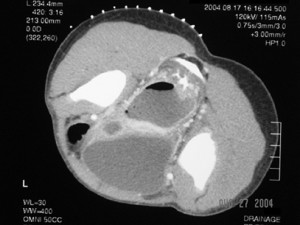
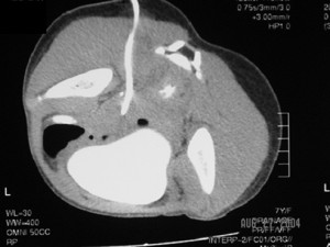
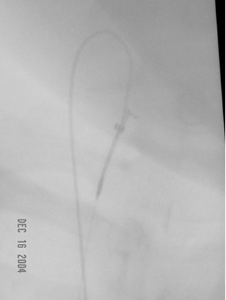
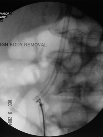
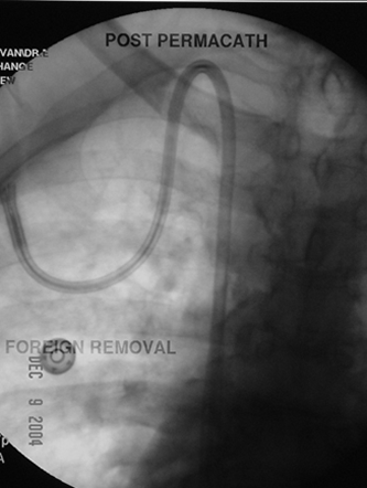
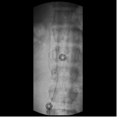
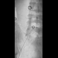
The top-row images belong to a middle-aged woman with skin metastasis from small cell lung cancer who developed a deep pelvic abscess. The first image shows planning of transgluteal drainage of the collection, while the second image captures the final position of a 12F pigtail catheter deployed into it.

CT-guided_drainage_pelvic abscess_1 CT-guided_drainage_pelvic abscess_2

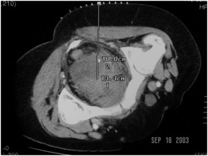
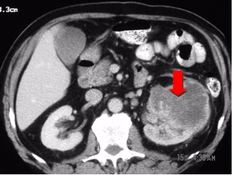
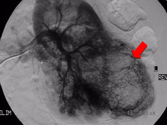
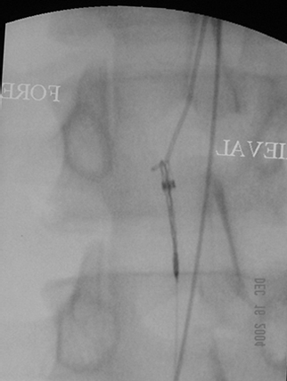
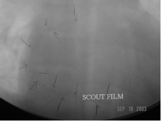
In the bottom row the first image illustrates the necessity of avoiding a frontal percutaneous approach to draining deep pelvic abscesses: this abscess is sandwiched between the rectum and the urinary bladder and in front of the bladder lies a loop of bowel and to its sides are the iliac vessels. The second image shows complete drainage of the abscess through the left buttock.

CT-guided_drainage_pelvic abscess_3 CT-guided_drainage_pelvic abscess_4

Embolization of left renal cell carcinoma prior to surgical removal. Ken U. Ekechukwu, MD, MPH, FACP.

Some tumors like renal cell carcinoma are very vascular and their surgical resection can be bloody. Some surgeons electively embolize such tumors before surgery to reduce intra-operative bleeding. 

This patient had carcinoma of his left kidney and I embolized it with dry alcohol and gel-foam before it was removed the next day. The first image is a contrast-enhanced transverse CT scan of the tumor showing a large deformed left kidney and the second image, angiogram of the tumor before I embolized it. The third image is angiogram of the left renal artery after embolization showing lack of opacification of the kidney.

Renal_carcinoma_1 Renal_carcinoma_2 Renal_carcinoma_3

Case 1: Retrieval of a lost guidewire from the IVC and right atrium. Ken U. Ekechukwu, MD, MPH, FACP.

Guidewires can disappear into patients during endovascular interventions such as the insertion of a central venous access. This typically occurs when the operator advances the guidewire and the dilator, sheath, or catheter as a unit, rather than advance these devices over the guidewire. The guidewire is simply a ‘guiding’ tool and should be regarded and employed as such; it is the stem upon which the interventionalist’s tubes and catheters glide to their destination. The wire should be fixed in position as the guided object is slid over it. The images below show the case of a patient from an emergency department in whom a guidewire disappeared into his right common femoral vein during a line placement. It was a 3mm J 0.035 inch guidewire and extended from the groin to the right atrium, but was safely retrieved with a snaring device passed up the inferior vena cava (IVC) from the contralateral groin into the right atrium. In the first image from the left, the guidewire is snared in the right atrium; in the second image, the snare and the wire are pulled into the IVC; the last image is a shot of the retrieved wire (shaped like a shepherd’s hook) and the snaring device (the wire with 3 loops at its end sticking out of a sheath).  

Retrieval_of_a_guidewire_1 Retrieval_of_a_guidewire_2 Retrieval_of_a_guidewire_3

Case 2: Retrieval of a cut central venous catheter from the right atrium. Ken U. Ekechukwu, MD, MPH, FACP.

Foreign bodies such as guidewires and catheters sometimes break off their parent body and travel through the blood stream to another site. This can happen in the process of removing them from or inserting them into the body. When it happens the embolized pieced should be removed from the body if it is safe to do so.

This patient had a dialysis unit composed of two large tunneled hemodialysis catheters inserted into his right atrium and attached to two subcutaneous chest ports. The unit malfunctioned and had to be replaced with a composite dual-lumen dialysis catheter. During their removal, however, one of the catheters was unintentionally cut and its leading piece, irretrievable by blunt tissue dissection, had to be fished from the patient with a snare that I introduced into his right atrium through his right common femoral vein.

The first image below shows the initial dialysis unit before removal; the second is an image of the snaring and retrieval process; while the third is a final image of the new dual-lumen dialysis catheter after its insertion.

Retrieval_of_central_venous_catheter_1 Retrieval_of_central_venous_catheter_2 Retrieval_of_central_venous_catheter_3

Case 3: Retrieval of a broken catheter from the right atrium. Ken U. Ekechukwu, MD, MPH, FACP.

Foreign bodies such as guide wires and catheters sometimes break off and travel through the blood stream from where they were placed originally to another site. When this happens they should be removed from the body if it is safe to do so. This man had the leading end of his central venous catheter pinched off by his right clavicle (or collar bone). The free piece of the catheter dropped into the right atrium and folded up there.

I ran a retrieval set from his right groin to his right atrium, snared the catheter and removed it. He did well afterwards.

Retrieval_of_foreign_bodies_1 Retrieval_of_foreign_bodies_2 Retrieval_of_foreign_bodies_3